| Home | E-Submission | Sitemap | Contact Us |  
J Epilepsy Res > Volume 3(2); 2013 > Article
Lee and Kim: Focal Cortical Dysplasia and Epilepsy Surgery

Abstract

Focal cortical dysplasia (FCD) is the most commonly encountered developmental malformation that causes refractory epilepsy. With advances in neuroimaging techniques, in particular MRI, recent studies have revealed a higher prevalence of FCD than previously estimated and have improved the preoperative identification and classification of these abnormalities. However, MRI frequently does not show any abnormalities in patients with pathologically proven FCD. In this situation, functional neuroimaing such as FDG-PET and ictal SPECT can be helpful. FCD is thought to be intrinsically epileptogenic, because the dysplastic tissues contain aberrant neural networks that are highly susceptible to abnormal excitation. The response to the medical treatment of epilepsy has been documented as consistently poor. Therefore, surgical resection has been an important alternative treatment for patients with intractable epilepsy related to FCD. Incomplete resection of FCD has been consistently known to be a poor prognostic factor. However, the complete removal of FCD is often difficult because the demarcation of the lesion is frequently poor, and dysplastic tissues tend to be more extensive than is apparent on MRI. Evidence indicates that even patients with MRI abnormalities who have resective epilepsy surgery for FCD have worse surgical outcomes than those of patients who have surgery for other focal lesional epilepsy syndromes. Careful planning of evelauation using intracranial electrodes is necessary for successful epilepsy surgery.

Terminology, incidence, and classification

Focal cortical dysplasia (FCD) is the most commonly encountered developmental malformation causing refractory epilepsy. Malformation of cortical development (MCD) is malformative lesions of the brain resulting from developmental aberration of normal processes that take place mostly during the first two trimesters of pregnancy and involving cells participating in the formation of the normal cerebral cortex. Neuronal migration disorder is regarded as an old terminology because not all MCDs are caused by neuronal migration anomaly. There are heterogeneous groups of focal or diffuse malformations depending on the causes and timing of the defect in the developmental processes.
The incidence of MCDs is variable according to the development and application of high resolution MRI. There is increasing tendency in the incidence of MCDs because of the advancement of this technique.1,2 It is the most common etiology of the cryptogenic and probable symptomatic epilepsy. It comprises 25 to 40% of the refractory childhood epilepsies.3 75% of patients with MCDs will have epilepsy at some time in their life.4
Both genetic and environmental factors play in the genesis of MCDs (Table 1)5. Recently, whole exom sequencing demonstrated that mutations of WD repeat domain 62 was the cause of wide spectrum of severe cortical malformation including microcephlay, pachygyria, lissencphaly, schizencephaly, polymicrogyria, and hypoplasia of corpus callosum.6 Another whole exom sequencing also identified de novo somatic mutations of PIK3CA, AKT3, and MTOR genes in focal brain areas.7
There is no general consensus on the classification of MCD because a particular defect in the cortical development may be responsible for more than one morphological subcategory of MCDs and one morphological subtype of MCD may arise from more than one mechanism for its formation. The most commonly used classification is Barkovich classification.8 It has four categories based on the major stages of brain development. They are malformation due to abnormal neuronal and glial proliferation, malformation due to abnormal neuronal migration, malformation due to abnormal cortical organization including later neuronal migration, and unclassified malformations. However, with the application of this system on the classification of FCD, FCD should be subdivided into two subcategories. FCD without balloon cells belongs to the category of malformation due to abnormal cortical organization including later neuronal migration and FCD with balloon cells belongs to the category of abnormal neuronal and glial proliferation.
FCD has intrinsic epileptogenicity because of the abnormal arrangement of the intralesional circuitry. The epileptogenic properties of dysmorphic neurons are well demonstrated in FCD. Ictal discharges and interictal spikes originated from FCD area where dysmorphic neurons were located.9 Seizures also started from the cortical area characterized by dysmorphic neurons but not balloon cells.10 Cytomegalic neurons demonstrated abnormal electrophysiological properties.11 There is imbalance between glutamergic excitatory and GABAergic inhibitory inputs.12,13 NR2A and NR2B subunits are increased in FCD area.14
Since the first distinctive and characteristic focal pathology was described,15 numerous classification schemes have been presented. Tassi et al16 proposed a new simplified classification based on the pathological characteristics with some other aspects including MRI findings and clinical considerations. They are architectural dysplasia, cytoarcitecural dysplasia, and Taylor-type cortical dysplasia. The common feature of these three entities was cortical laminar disruption. The distinction was based on the presence of cytological abnormalities such as giant neurons, dysmorphic neurons, and balloon cells.
Palmini et al17 further refined the terminology of classification of FCD with continuing criteria of histopatholgical findings. Recently ILAE proposed the new classification.18 In this system, FCD was classified into three entities (Table 2). The whole mark of Type I FCD is the cortical dyslamination and that of type II is the presence of dysmorphic neuron with or without balloon cells in addition to the cortical dyslamination. If there is another lesion other than type I or type II pathology, it is categorized into FCD type III.

Neuroimaging findings

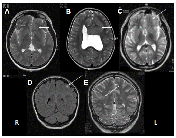
MRI features of FCD are variable. They are focal increase of cortical thickness, blurring of gray-white matter junction, increased signal in T2-weighted or FLAIR imaging, and increased signal from the cortical surface to the ventricle (Fig. 1). The final characteristic finding is called transmantle sign. Thin slice section and concomitant use of T2 and FLAIR imaging are important in detecting small FCD. The diagnostic sensitivity is increased along with the severity of pathology of FCD (Table 3).19 Thirty-seven percent of the patients with type I FCD had normal MRI while only 15% with FCD type II had negative MRI.16,1922 The thick gray matter, blurring gray and white matter junction, and increased T2 or FLAIR signal are more frequently associated with type II FCD pathology. However, Most MRI findings could not differentiate Type I from Type II pathology because of significant overlapping of imaging findings between FCD type I and type II.23 Cortical thickness and blurring of GW junction were more common in isolated FCD than in FCD type III. And the transmantle sign on MRI was more specific to Type II.
The severer pathology might mean the higher epileptogenicity in FCD. However, the most severe pathological presentation can be found in the patients with negative MRI. The proportion of negative MRI in the patients with pathologically proven FCD ranges from 23% to 73%.2426
Ictal SPECT and FDG-PET can be critical for localizing the seizure focus, especially in MR-negative patients. FDG-PET and subtraction SPECT are valuable in the diagnosis of MR-negative neocortical epilepsy.2732 For focal cortical dysplasia, the diagnostic sensitivity of FDG-PET and ictal SPECT ranges from 69% to 98% and 48% to 64%, respectively.21,3338

Epilepsy surgery of FCD

The proportion of FCD in surgical series ranges from 9% to 71% depending on the slection criteria of different centers.16,19,22,3436,3942 Our series40 included only neocortical epilepsy patients. In this series the proportion of FCD reached 71%. The relative poor outcome of FCD compared with benign brain tumor or temporal lobe epilepsy (TLE) with hippocampal sclerosis can be noticed. Whereas approximately 80% of patients became seizure free after surgical treatment for mesial TLE related to HS or lesional epilepsy such as primary brain tumor or vascular malformations,43,44 the efficacy of surgical treatment for FCD was consistently less favorable, with approximately 33–75% of individuals becoming seizure free.16,36,39,45 The relative poor prognosis can be explained by that FCD is often invisible on MRI which leads to the difficulty to define epileptogenic zone. We have to depend only on semiology, EEG, and functional neuroimaging. The other factor affecting the relative poor surgical outcome is that even after the removal of all visible lesions on MRI, residual microscopic lesion can still be epilepstogenic, which means that dysplastic tissues tend to be more extensive than is apparent on MRI.21,46 Evidence indicates that even patients with MRI abnormalities who have resective epilepsy surgery for FCD have worse surgical outcomes than those of patients who have surgery for other focal lesional epilepsy syndromes.40 The complete resection of FCD is invariably associated with better surgical outcome (Table 4).19,20,22,36
Although MRI increases our confidence in the localization of epileptogenic foci and allows surgical resection to be performed without preoperative invasive studies, our series47 demonstrated that complete resection guided by areas of ictal onset, persistent pathological delta slowing, >1/sec frequent spikes, and intermittent fast activity (HFO) detected by intracranial EEG was very useful leading to the complete resection even in the patients with negative MRI. The importance of complete resection indicates that seizures may originate from periphery of FCD as well as the center of FCD. There is an inherent difficulty in identifying the epileptogenic zone in MR-negative FCD patients, which often results in incomplete resection. However, with careful interpretation of other studies including functional neuroimaging and concordant results among these studies, surgical treatment can benefit patients with MR-negative FCD.
The positive prognostic effect of severe pathological features has been documented in previous reports, which predominantly included patients with identifiable lesions on MRI.2,16,45,48 Our series19 also identified that the patients with severe pathological features have a higher chance of becoming seizure free after surgery regardless of MRI results. The prognostic role of pathological severity may be related to the completeness of surgical resection. Although the epileptogenic zone in FCD does not always correspond to the area with the most severe pathological features, it is generally accepted that the epileptogenicity of FCD is usually associated with its pathological severity. From this point of view, the severe pathology may mean more confined and higher epileptogenic area. The severe pathology can also easily be identified by other neuroimaging techniques. Furthermore, mild and severe pathology may have different characteristics of epileptogenic network. In other words, numerous variable entities may be clustered in mild pathology.

Conclusions

FCD is the most common MCD which is a major cause of epilepsy. Most MCDs are epileptogenic: FCD has intrinsic epileptogenicity. Neuroimaging development, especially high resolution MRI, can increase the detection rate of MCD, which can make us treat the patients surgically. FDG-PET and ictal SPECT can be valuable in the localization of FCD, which may be especially useful in the patients with normal MRI. Surgical treatment is a option for refractory epilepsy with FCD and complete resection of FCD area is the major prognostic factor. Complete resection of early ictal onset zone and resection which includes the area with pathological delta waves and frequent interictal spikes predict good surgical outcome. Pathologic characteristics are important not only for the correct diagnosis but also for prediction of surgical prognosis.

Figure 1.
MRI findings of focal cortical dysplasia. (A, B) Thick gyri (arrows), (C) Blurring of cortico-white matter junction, (D) Focal high signal, (E) Transmantle sign.
er-3-2-43-1f1.gif
Table 1.
The type of MCDs and the possible related gene defects
Type of MCD Possible related gene defects
FCD, Cortical tuber TSC1, TSC2 (controversial)
Polymicrogyria SPRX2, KIAA1279, GPR56, PAX6, TBR2, COL18A1, RABG3GAP1, TUB2B, 22q11.2
Periventricular nodular heterotopia (PNH) FLNA1, ARFGEF2, LRP2, Various copy number variation
Subcortical band heterotopia DCX,LIS1
Lissencephaly LIS1, DCX, Microdeletion in 17p, ARX, TUBA1A, RELN
Schizencephaly EMX2
Table 2.
Classification of FCD
FCD Type I FCD Type II FCD Type III
Type IA Type IIA Type IIIA
FCD with abnormal radial cortical lamination FCD with dysmorphic neurons Cortical dyslamination associated with HS
Type IB Type IIB Type IIIB
FCD with abnormal tangential cortical lamination FCD with dysmorphic neurons and balloon cells Cortical dyslamination associted with glial and glioneuronal tumor
Type IC Type IIIC
FCD with abnormal radial and tangential lamination Cortical dyslamination adjacent to vascular malformation
Type IIID
Cortical dyslamination adjacent other acquired lesions
Table 3.
Diagnostic sensitivity of MRI and pathology of FCD
MRI mMCD FCD 1A FCD 1B FCD 2A FCD 2B p-value
Diagnostic sensitivity 19.0% (4/21) 30.3% (27/89) 60.7% (17/28) 75.0% (9/12) 81.3% (13/16) <0.001
False negative rate, % 81.0 69.7 39.3 25.0 18.7
Table 4.
The relationship between the surgical outcome and complete resection
Study Complete resection Incomplete resection
Kloss et al.20 (2002) 80% 17%
Kim et al.19 (2009) 82% 30%
Krsek et al.22 (2009) 70% 22%
Alexandre et al.36 (2006) 86% 50%

References

1. Kuzniecky RJ, Jaclson GD. Magnetic resonance imaging in epilepsy: Neuroimaging techniques. 2nd ed. New York: Elsevier; 2005.

2. Widdess-Walsh P, Diehl B, Najm I. Neuroimaging of focal cortical dysplasia. J Neuroimaging. 2006;16:185–96.
crossref pmid
3. Guerrini R, Holthausen H, Pameggiani L, et al. Epilepsy and malformations of the cerebral cortex. Roger J, Bureau M, Dravet C, editors. Epilepticc syndromes in infancy, childhood and adolescence. 3rd Ed. London: John Libbey; 2002. p. 457–479.
crossref pmid
4. Leventer RJ, Phelan EM, Coleman LT, et al. Clinical and imaging features of cortical malformations in childhood. Neurology. 1999;53:715–722.
crossref pmid
5. Spreafico R, Tassi L. Cortical malformations. Stefan H, Theodre W H, editors. Handbook of clinical neurology vol 108(3rd series): Epilepsy part II. Amsterdam: Elsevier; 2012. p. 536–557.
crossref pmid
6. Bilgüvar K, Oztürk AK, Louvi A, et al. Whole-exome sequencing identifies recessive WDR62 mutations in severe brain malformations. Nature. 2010;467:207–10.
crossref pmid pmc
7. Lee JH, Huynh M, Silhavy JL, et al. De novo somatic mutations in components of the PI3K-AKT3-mTOR pathway cause hemimegalencephaly. Nature Genet. 2012;44:941–5.
crossref pmid
8. Barkovich AJ, Kuzniecky RJ, Jackson GD, et al. A developmental and genetic classification for malformations of cortical development. Neurology. 2005;65:1873–87.
crossref pmid
9. Chassoux F, Devaux B, Landré E, et al. Stereoelectroencephalography in focal cortical dysplasia: a 3D approach to delineating the dysplastic cortex. Brain. 2000;123:1733–51.
crossref pmid
10. Boonyapisit K, Najm I, Klem G, et al. Epileptogenicity of focal malformations due to abnormal cortical development: direct electrocorticographic-histopathologic correlations. Epilepsia. 2003;44:69–76.
crossref
11. Cepeda C, Hurst RS, Flores-Hernández J, et al. Morphological and electrophysiological characterization of abnormal cell types in pediatric cortical dysplasia. J Neurosci Res. 2003;72:472–86.
crossref pmid
12. Mikuni N, Babb TL, Ying Z, et al. NMDA-receptors 1 and 2A/B coassembly increased in human epileptic focal cortical dysplasia. Epilepsia. 1999;40:1683–7.
crossref pmid
13. Ying Z, Babb TL, Mikuni N. Selective coexpression of NMDAR2A/B and NMDAR1 subunit proteins in dysplastic neurons of human epileptic cortex. Exp Neurol. 1999;159:409–18.
crossref pmid
14. White R, Hua Y, Scheithauer , et al. Selective alterations in glutamate and GABA receptor subunit mRNA expression in dysplastic neurons and giant cells of cortical tubers. Ann Neurol. 2001;49:67–78.
crossref pmid
15. Taylor DC, Falconer MA, Bruton CJ, et al. Focal dysplasia of the cerebral cortex in epilepsy. J Neurol Neuropsychiatry. 1971;34:369–87.
crossref pmid pmc
16. Tassi L, Colombo N, Garbelli R, et al. Focal cortical dysplasia: neuropathological subtypes, EEG, neuroimaging and surgical outcome. Brain. 2002;125:1719–32.
crossref pmid
17. Palmini A, Najim I, Avanzini G, et al. Terminology and classification of the cortical dysplasia. Neurology. 2004;62:2–8.
crossref pmid
18. Blumcke I, Thom M, Aronica E, et al. The clinicopathologic spectrum of focal cortical dysplasia: a conseneus classification propsed by an ad hoc Task Force of the ILAE Diagnostic Methods Commission (1). Epilepsia. 2011;52:158–74.
crossref pmid pmc
19. Kim DW, Lee SK, Chu K, et al. Predictors of surgical outcome and pathologic considerations in focal cortical dysplasia. Neurology. 2009;72:211–216.
crossref pmid
20. Kloss S, Pieper T, Pannek H, et al. Epilepsy surgery in children with focal cortical dysplasia (FCD): results of long-term seizure outcome. Neuropediatrics. 2002;33:21–6.
crossref pmid
21. Cohen-Gadol AA, Ozduman K, Bronen RA, et al. Long-term outcome after epilepsy surgery for focal cortical dysplasia. J Neurosurg. 2004;101:55–65.
crossref pmid
22. Krsek P, Maton B, Jayakar P, et al. Incomplete resection of focal cortical dysplasia is the main predictor of poor postsurgical outcome. Neurolory. 2009;72:217–23.
crossref pmid
23. Kim DW, Kim S, Park S, et al. Comparison of MRI features and surgical outcome among the subtypes of focal cortical dysplasia. Seizure. 2012;21:789–94.
crossref pmid
24. Chapman K, Wyllie E, Najm I, et al. Seizure outcome after epilepsy surgery in patients with normal preoperative MRI. J Neurol Neurosurg Psychiatry. 2005;76:710–3.
crossref pmid pmc
25. Lee SK, Lee SY, Kim KK, Hong KS, Lee DS, Chung CK. Surgical outcome and prognostic factors of cryptogenic neocortical epilepsy. Ann Neurol. 2005;58:525–32.
crossref pmid
26. McGonigal A, Bartolomei F, Régis J, et al. Stereoelectroencephalography in presurgical assessment of MRI-negative epilepsy. Brain. 2007;130:3169–83.
crossref pmid
27. Geier S, Bancaud J, Talairach J, Bonis A, Szikla G, Enjelvin M. The seizures of frontal lobe epilepsy. Neurology. 1977;27:951–8.
crossref pmid
28. Chugani HT, Shewmon DA, Shields WD, et al. Surgery for intractable infantile spasms: neuroimaging perspectives. Epilepsia. 1993;34:764–771.
crossref pmid
29. Duncan R, Biraben A, Patterson J, et al. Ictal single photon emission computed tomography in occipital lobe seizures. Epilepsia. 1997;38:839–43.
crossref pmid
30. Henry TR, Babb TL, Engel J, Mazziotta JC, Phelps ME, Crandall PM. Hippocampal neuronal loss and regional hypometabolism in temporal lobe epilepsy. Ann Neurol. 1994;36:925–7.
crossref pmid
31. Shields WD, Duchowny MS, Holmes GL. Surgically remediable syndromes of infancy and early childhood. Engel J, editor. Surgical treatment of the epilepsies. 2nd ed. New York: Raven Press; 1993. p. 35–48.

32. Williamson PD, Spencer DD, Spencer SS, Novelly RA, Mattson RH. Complex partial seizures of frontal lobe origin. Ann Neurol. 1985;18:497–504.
crossref pmid
33. Hader WJ, Mackay M, Otsubo H, et al. Cortical dysplastic lesions in children with intractable epilepsy: role of complete resection. J Neurosurg. 2004;100:2 Suppl Pediatrics. 110–7.
crossref pmid
34. Lerner JT, Salamon N, Hauptman JS, et al. Assessment and surgical outcomes for mild type I and severe type II cortical dysplasia: a critical review and the UCLA experience. Epilepsia. 2009;50:1310–35.
crossref pmid
35. Park CK, Kim SK, Wang KC, et al. Surgical outcome and prognostic factors of pediatric epilepsy caused by cortical dysplasia. Childs Nerv Syst. 2006;22:586–92.
crossref pmid
36. Alexandre V, Walz R, Bianchin MM, et al. Seizure outcome after surgery for epilepsy due to focal cortical dysplastic lesions. Seizure. 2006;420–7.
crossref pmid
37. Gupta A, Raja S, Kotagal P, et al. Ictal SPECT in children with partial epilepsy due to focal cortical dysplasia. Pediatr Neurol. 2004;31:89–95.
crossref pmid
38. Kim SK, Na DG, Byun HS, et al. Focal cortical dysplasia: comparison of MRI and FDG-PET. J Comput Assist Tomogr. 2000;24:296–302.
crossref pmid
39. Siegel AM, Cascino GD, Meyer FB, Marsh WR, Scheithauer BW, Sharbrough FW. Surgical outcome and predictive factors in adult patients with intractable epilepsy and focal cortical dysplasia. Acta Neurol Scand. 2006;113:65–71.
crossref pmid
40. Yun CH, Les SK, Lee SY, et al. Prognostic factors in neocortical epilepsy surgery: multivariate analysis. Epilepsia. 2006;47:574–9.
crossref pmid
41. Fauser S, Schulze-Bonhage A, Honegger J, et al. Focal cortical dysplasias: surgical outcome in 67 patients in relation to histological subtypes and dual pathology. Brain. 2004;127:2406–18.
crossref pmid
42. Kral T, von Lehe M, Podlogar M. Focal cortical dysplasia: long term seizure outcome after surgical treatment. J Neurol Neurosurg Psychiatry. 2007;78:853–6.
crossref pmid pmc
43. Britton JW, Cascino GD, Sharbrough FW, Kelly PJ. Low-grade glial neoplasms and intractable partial epilepsy: efficacy of surgical treatment. Epilepsia. 1994;35:1130–5.
crossref pmid
44. Jeong SW, Lee SK, Hong KS, Kim KK, Chung CK, Kim H. Prognostic factors for the surgery for mesial temporal lobe epilepsy: longitudinal analysis. Epilepsia. 2005;46:1273–9.
crossref pmid
45. Chung CK, Lee SK, Kim KJ. Surgical outcome of epilepsy caused by cortical dysplasia. Epilepsia. 2005;46:Suppl 1. 25–9.
crossref pmid
46. Sisodiya SM, Free SL, Stevens JM, Fish DR, Shorvon SD. Widespread cerebral structural changes in patients with cortical dysgenesis and epilepsy. Brain. 1995;118:1039–50.
crossref pmid
47. Kim DW, Kim HK, Lee SK, et al. Extent of neocortical resection and surgical outcome of epilepsy: Intracranial EEG analysis. Epilepsia. 2010;51:1010–7.
crossref pmid
48. Hardiman O, Burke T, Phillips J, et al. Microdysgenesis in resected temporal neocortex: incidence and clinical significance in focal epilepsy. Neurology. 1988;38:1041–7.
crossref pmid
TOOLS
PDF Links  PDF Links
PubReader  PubReader
XML Download  XML Download
Full text via DOI  Full text via DOI
Download Citation  Download Citation
  Print
Share:      
METRICS
31
Crossref
28,023
View
197
Download
Editorial Office
KCC Parktown 101-308, Mallijae-ro 185, Jung-Gu, Seoul, Korea
TEL : +82-362-9662   FAX : +82-2-362-9663   E-mail : epilepsy@kes.or.kr

Copyright© Korean Epilepsy Society.              Developed in M2PI
About |  Browse Articles |  Current Issue |  For Authors and Reviewers