Introduction
Cognitive dysfunction is frequently observed in epilepsy patients. As antiepileptic drugs (AEDs) are commonly taken for extended periods of time, the chronic use of AEDs may be a cause of cognitive dysfunction in this population.1–3 Thus, the impact of AED-related cognitive impairment on daily life is an important issue to consider in the treatment of epilepsy.
Oxcarbazepine (OXC) is a second generation AED that is chemically related to carbamazepine (CBZ) and approved as initial or add-on therapy for epilepsy patients. Neuropsychological (NP) tests show that, compared to traditional AEDs, OXC is not associated with significant cognitive side effects in both epilepsy patients and healthy subjects.2 While NP tests are a widely used method for detecting the effect of AEDs on cognitive function, there may be a significant test-retest variability and practice effect from repeated testing.4 Furthermore, NP tests cannot gather temporal information about cognitive processing by the brain.
Spectral analysis is based on changes in the electroencephalogram (EEG) spectrum, which reflects the functional state of the brain. Spectral analysis of electroencephalographic signals constitutes a helpful tool in the assessment of pharmacological effects of CNS drugs, such as AEDs, on the brain. It has proven to be a valuable technique for classifying psychopharmacological agents and assessing their pharmacodynamics. Therefore, it can serve as a simple and objective method to assess the relationship between AEDs and cognitive function.5–7 However, spectral EEG reflects only a general functional state of the brain including cognitive function. Given this drawback, NP tests are frequently necessary for proper interpretation of the spectral EEG findings.
To date, only a few studies examining OXC effects on spectral EEG have been reported in epilepsy patients.8,9 Clemens et al. reported OXC effects on spectral EEG in nine patients with epilepsy (mean age 18.8 years).8 They demonstrated that OXC had no significant effect on the spectral power in broad bands. Another study showed that OXC treatment was characterized by less delta, theta, and alpha power, and more beta power than carbamazepine treatment.9 However, as the study did not perform NP tests in conjunction with spectral EEG, the cognitive effects of OXC may be subject to misinterpretation.
In the present study, we investigated the effects of OXC on cognitive function using both NP tests and spectral power in an attempt to gather more useful information on cognitive impairment.5 We also adopted spectral coherence as a tool for identifying functional connectivity, given its unique ability to reveal synchronous interactions between two brain locations, which cannot be performed by the NP test or spectral EEG.10 Recently, spectral coherence approaches have been applied to observe functional connectivity in Alzheimer disease,11 epilepsy,12 alcoholism,13 and chronic fatigue syndrome.14
Material and Methods
Subject selection and OXC administration
Inclusion criteria were as follows. 1) All subjects had a newly diagnosed epilepsy with at least 2 unprovoked partial or secondarily generalized seizures without previous exposure to AEDs. The diagnosis of epilepsy was made based on the revised classification of epilepsies and epileptic syndromes.15 2) The age of subjects limited to between 18 and 55 years. 3) Subjects had received EEG and NP tests twice, both before and after OXC treatment. Exclusion criteria included; 1) seizures evoked by metabolic abnormality, toxic exposure, active infection, or other treatable causes, 2) special seizure types such as absence, myoclonic, or atonic seizures, 3) use of acute anticonvulsant medications longer than one week, use of other central nervous system (CNS) acting drugs, 4) a diagnosis of mental retardation, progressive neurological disease or psychiatric disease, 5) pregnant or breat-feeding women or women who wanted to have baby within a year, 6) history of status epilepticus, and 7) abnormal laboratory findings including neutrophil count less than 1,800/mm3, platelet count less than 100,000/mm3, twice higher than upper normal range of aspartate aminotransferase (AST), alkaline aminotransferase (ALT), bilirubin, blood urea nitrogen (BUN), creatinine, sodium, and potassium levels. Fifteen patients met these criteria and were included in the final analysis.
Before OXC treatment, all subjects underwent a physical examination, routine blood tests including hematology and liver function tests, magnetic resonance imaging (MRI) of the brain, EEG, and NP tests. All tests were performed both at the study onset and 35 weeks (35.5± 6.39) post-drug initiation. OXC was given at 300 mg a day for the first two weeks, 600 mg a day for the next two weeks, then maintained with the same dosage. If seizures persisted after the initial target of dose 600 mg/day, the dosage was increased up to 1800 mg/dayuntil seizures were controlled. If a patient noted any intolerable side effects from OXC administration, the dosage was decreased by 300 mg a day for one or 2 weeks until symptoms subsided. A fixed doses of OXC were maintained for at least four weeks prior to conducting the second NP test and EEG study.
The frequency of seizures was assessed using a self-recorded seizure diaries brought in by subjects at subsequent outpatient clinic visits. Informed consent was obtained from all subjects in accordance with the institutional review board’s guidelines at each hospital participating in the study.
EEG recording
In accordance with the international 10–20 system, EEGs were recorded using Ag/AgCl electrodes that were placed at Fp1, Fp2, F7, F3, Fz, F4, F8, T3, C3, Cz, C4, T4, T5, P3, Pz, P4, T6, O1, and O2. Electrode impedance was kept below 5 kΩ, and the band-pass filter (0.5–70 Hz) was applied. The sampling rate was 200–400 Hz. The reference electrode was set to Pz electrode. Two electrooculography (EOG) channels (placed on the left and right outer canthi) were added to confirm eyeball movements and eliminate EOG artifacts. Subjects rested supine in a bed during EEG recordings and were asked to close their eyes for 10s and then open for another 10s. This procedure was repeated 10 times.
EEG data preprocessing and analysis
Epoch selection
For quantitative analysis of background EEG activity, an epilepsy specialist (JKY) selected epochs while blinded to the demographic data in our study. Epochs with artifacts or with epileptiform activities (spikes or sharp waves) were eliminated, while epochs with physiological alpha activity of maximum amplitude in occipital regions (O1 and O2 electrode) were selected. We collected 20 1.5 s-long epochs in each patient before and after OXC treatment.
Data preprocessing
EEG data was preprocessed using EEGLAB version 10.0b16 managed in the MATLAB environment (version 7.10, The Mathworks, Natick, MA, USA). After down-sampling to 200 Hz, EEG data were re-referenced to the common average reference, and band pass filtered (0.5–50 Hz) for analysis.
Spectral power
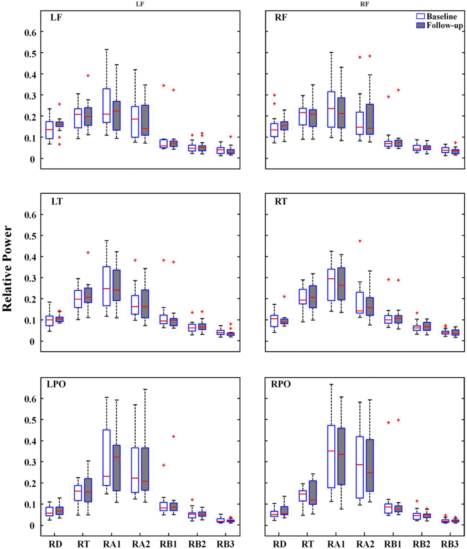
To identify spectral power, fast Fourier transform was applied to each of the selected epochs in the first step. Then, to improve the accuracy of spectral estimates all transformed epochs for each subject were averaged. Frequency band of 1 to 30 Hz with 0.4 Hz steps was used, and were then divided into seven discrete bands: Delta: 1–3 Hz, Theta: 3–7 Hz, Alpha 1: 7–10 Hz, Alpha 2: 10–13 Hz, Beta 1: 13–19 Hz, Beta 2: 19–24 Hz, and Beta 3: 24–30 Hz. In order to obtain relative powers, absolute powers of each frequency bin divided by sum of the overall spectral powers of each subject. Relative powers were log-transformed prior to statistical analysis. The electrodes were grouped into six scalp regions: LF (left frontal: Fp1, F7, F3), RF (right frontal: Fp2, F8, F4), LT (left temporal: T7, C3), RT (right temporal: T8, C4), LPO (left parieto-occipital: P3, P7, O1), and RPO (right parie-to-occipital: P4, P8, O2).
Spectral Coherence
To calculate spectral coherence, magnitude square coherence (the mscohere function in signal processing toolbox of MATLAB) was used. Hanning windows were successively applied to 1.5s epochs of EEG with 50% overlap. Spectral coherence Cxy between two brain signals x(t) and y(t) was defined as
where Cxy was the cross-spectral density between x and y, and Gxx and Gyy were the autospectral densities of x and y, respectively. For spectral coherence calculation, spectral power ranged from 1 to 30 Hz and the interval was 0.4 Hz. Intrahemispheric spectral coherences were calculated for the eight electrode pairs (F3-C3, F4-C4, T3-T5, T4-T6, C3-P3, C4-P4, P3-O1 and P4-O2), and interhemispheric spectral coherences were computed for the six electrode pairs (F3-F4, T3-T4, T5-T6, C3-C4, P3-P4 and O1-O2) in each of the six discrete frequency bands.10
Neuropsychological assessments
A series of NP tests were selected to investigate cognitive functions including attention, language, visuospatial abilities, motor skills, memory, and executive functions of the frontal lobes. The digit span test forward was administered to assess attention.17 Language was evaluated using the the short version of the Korean-Boston naming test (S-K-BNT) form A.18 Visuo-spatial function was evaluated using the Rey complex figure test (RCFT).19 Motor function was evaluated using the finger tapping test (FTT).20 To assess memory, we used two sub-tests: the Korean-California verbal learning test (K-CVLT)21 and the RCFT.19 To investigate the executive functions of the frontal lobe, we used five sub-tests: the controlled word association task (COWAT),22 the Korean-color word stroop test (K-CWST),23 the digit symbol test,24,25 the trail making test (TMT),26 and the Wisconsin card sorting test (WCST).27
Statistical analysis
Wilcoxon signed-rank tests were used to compare patients’s grand averages power spectra within each frequency bands, and NP test scores before and after the initiation of medication for each subject. Repeated measures analysis of variance (ANOVA) was used to analyze spectral powers of ROIs. The within-subject factors included treatment (two levels: baseline and follow-up) and electrode region (six levels: LF, RF, LT, RT, LPO, and RPO). Included among within-subject factors were cases of spectral coherence: electrode pair (eight levels for intrahemispheric spectral coherence, six levels for in-terhemispheric spectral coherence). The Greenhouse-Geisser correction was used to evaluate F ratios to control for type 1 errors in the repeated measures design. The significance level was set to 0.05. All statistical procedures were performed with SPSS (Version 12.0, Chicago, IL, USA).
Results
Subjects
Subject demographic data and clinical characteristics are presented in Table 1. Seven of the fifteen subjects were men, and the mean age of the patients was 30.7±10.3 years (range: 18–55 years). The mean interval between EEGs was 248.5±46.3 days (range: 206–348 days). The mean daily dosage of OXC was 740±365.1 mg (range: 300–1,800 mg).
Quantitative EEG analysis
Spectral analysis
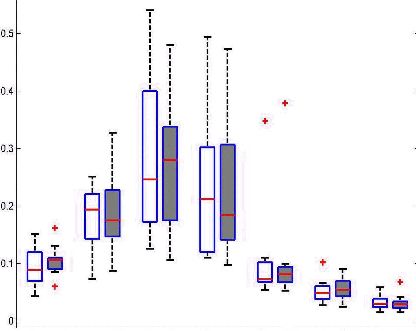
For spectral power of each frequency band across all electrodes, the signed-rank test revealed no significant difference between baseline and follow-up EEGs (Fig. 1). Repeated measures ANOVA revealed no significant main effect of treatment on the spectral power of each frequency band at the ROIs. Spectral power was significantly different across regions (p<0.005). However, the interaction between treatment and regions was not significant (Fig. 2).
Spectral coherence
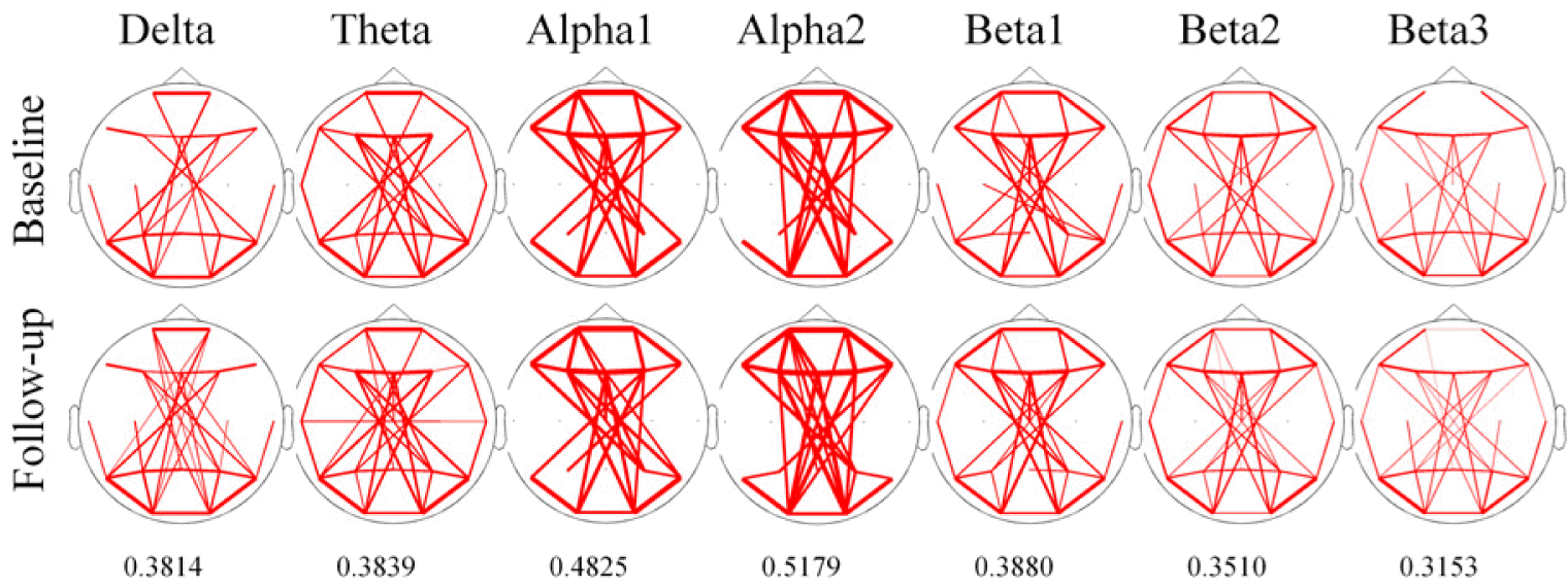
To identify overall pattern of coherence at each frequency band across all electrode pair, we draw the line topographic map (Fig. 3). Threshold of coherence were selected values as represented by mean+1 SD of coherence at each frequency bands. Line topography of coherence revealed that there were no difference of patterns after treatment in all frequency bands.
A statistical summary of spectral coherence is presented in Table 2. For intrahemispheric coherence, repeated measures ANOVA revealed a significant main effect of treatment in all frequency bands. However, the interaction between treatment and electrode pair did not reach statistical significance. For interhemispheric coherence, statistical results were the same as intrahemispheric coherence.
Neuropsychological tests
Statistical results of the NP tests are listed in Table 3. Significant differences were observed in the RCFT copy time (Z=−1.079, p=0.031), K-CVLT 1–5 trial total (Z=−1.929, p=0.054), digit symbol test (Z=−2.639, p=0.008), and TMT-B (Z=−1.988, p=0.047). The time to copy the Rey complex figure and to complete TMT-B was significantly decreased, while scores for the K-CVLT 1–5 trial total and digit symbol test were significantly increased. Significant differences were not noted in the other tests.
Discussion
In the present study, we investigated the effect of OXC monotherapy on background EEG spectral power, spectral coherence, and found that OXC was not associated with a significant change in spectral power or coherence; however, some cognitive functions improved. NP tests demonstrated improved performance in the copy trial of the RCFT (visuospatial function), K-CVLT (memory), digit symbol test, and TMT-B (executive function of frontal lobes). These findings are consistent with previous studies demonstrating OXC’s positive effects on a wide range of cognitive functions such as visuo-spatial organization,28,29 memory,30 focused attention, manual writing speed,31 and mental processing speed.29
No significant changes in frequency band powers were observed with OXC treatment, which is congruent with previous study.8 Although the effects of OXC on spectral EEG have been reported, the functional connection between brain regions remains unclear. Spectral coherence may be interpreted as the frequency-indexed correlation coefficient estimating the linear relationship between two time series. Thus, coherence means synchrony or coupling of two regions in a given frequency band.32 Neither intrahemispheric nor interhemispheric coherence changed significantly for all electrode pairs, indicating that OXC did not have a significant effect on the functional connectivity of EEG in our study.
Spectral coherence has been the main method used to assess the degree of functional connectivity between brain areas and associations between different functional states, however, the main limitation of this approach is that it investigates only a linear statistical link between EEG signals. Therefore, one cannot rule out the possibility of changes in nonlinear functional connectivity between brain regions following OXC treatment.33,34
AEDs can be categorized into two groups according to their effect on both spectral EEG and cognitive function (Table 4). Most older AEDs, including phenytoin35–37 and carbamazepine,8,9,37–41 as well as newer AEDs such as topiramate42–46 and gabapentine,40,45 tend to increase delta and theta power and decrease higher frequency bands. This first category of AEDs negatively affects cognitive function in patients with epilepsy. Increased EEG slowing is well correlated with cognitive decline in demented patients.47 In the case of carbamazepine and phenytoin, reported slowing of EEG background rhythm has been positively correlated with negative neuropsychological results as measured by cognitive testing.37,38,40,48
On the other hand, lamotrigine,8,43,46 levetiracetam5,39,49 and valproate8,50 are reported to reduce the power of slow frequencies and/or increase that of fast frequencies, thereby improving some cognitive function in diverse NP tests.2,51,52 In subjects treated with levetiracetam, the alpha and beta bands were positively correlated with improved neuropsychological results.5,53
Unlike AEDs of two categories above, OXC had no significant effect on spectral EEG, despite some cognitive functions were improved with OXC treatment. Thus, OXC seems to be an unique pharamacological effect on the brain in patients with epilepsy. However, as the number of patients was relatively small in the present study, larger sample of the study will be required to corroborate our study.
Brain activity during the resting state may not be captured on EEG compared with a cognitively functioning brain state that demonstrates changes in attention, inhibition, memory, and so forth. Moreover, it has been reported that there is no direct or causal relationship between EEG background measures and cognitive functions.7,49 This finding might suggest discrepancies between resting state spectral EEG and NP tests in our study. Accordingly, future studies should consider event-related potential study in addition to resting state EEG in order to better identify the effects of AEDs on cognitive function. In conclusion, our study supports the notion that OXC has no significant cognitive side effect in patients with epilepsy.