Issues of Women with Epilepsy and Suitable Antiseizure Drugs
Article information
Abstract
Seizure aggravation in women with epilepsy (WWE) tends to occur at two specific times during the menstrual cycle: the perimenstrual phase and the ovulation period. Antiseizure drugs (ASDs), especially those that induce enzymes, can accelerate the metabolism of hormones in oral contraceptives, rendering them less effective. Estrogen in contraceptive pills increases the metabolism of lamotrigine. Physiological changes during pregnancy can significantly impact the pharmacokinetics of ASDs, potentially necessitating adjustments in dosage for women with epilepsy to maintain seizure control. The use of valproate in pregnant women is associated with the highest risk of major congenital malformations among ASDs. Risks of major congenital malformations associated with lamotrigine, levetiracetam, and oxcarbazepine were within the range reported in the general population. Exposure to valproate can lead to lower IQ in offspring. Reduced folic acid levels are linked to orofacial clefts, cardiovascular malformations, and urogenital and limb anomalies in WWE. Decreased folate levels are expected with the use of enzyme-inducing ASDs. However, a high dose of folate was associated with an increased risk of cancer in children of mothers with epilepsy. Most ASDs are generally considered safe for breastfeeding and should be encouraged. However, no single ASD is considered ideal for childbearing WWE. Lamotrigine and levetiracetam are relatively more suitable options for this situation.
Introduction
Numerous factors require consideration when treating women with epilepsy (WWE), particularly those of childbearing potential. These factors encompass hormonal changes, fertility, pharmacokinetic alterations during pregnancy, and the effects of antiseizure drugs (ASDs) on the fetus. A thorough understanding of these matters is crucial for the appropriate management of this population.
Catamenial Epilepsy
The menstrual cycle involves a complex interplay of hormones that prepare the body for possible pregnancy, comprising three phases: the follicular (or proliferative) phase, ovulation, and the luteal (or secretory) phase. Commencing with the onset of menstruation and culminating with ovulation, the follicular phase sees rising estrogen levels as ovarian follicles mature. Estrogen contributes to the development of the uterine lining (endometrium). Approximately around day 14 in a 28-day cycle, a mature egg is released from the ovarian follicle, triggered by a surge in luteinizing hormone. This period coincides with peak estrogen levels. Post ovulation, the follicle becomes the corpus luteum, generating progesterone. Progesterone readies the endometrium for potential pregnancy. If pregnancy doesn’t transpire, the corpus luteum shrinks, prompting a decline in progesterone and initiating menstruation. This signifies the luteal phase.1
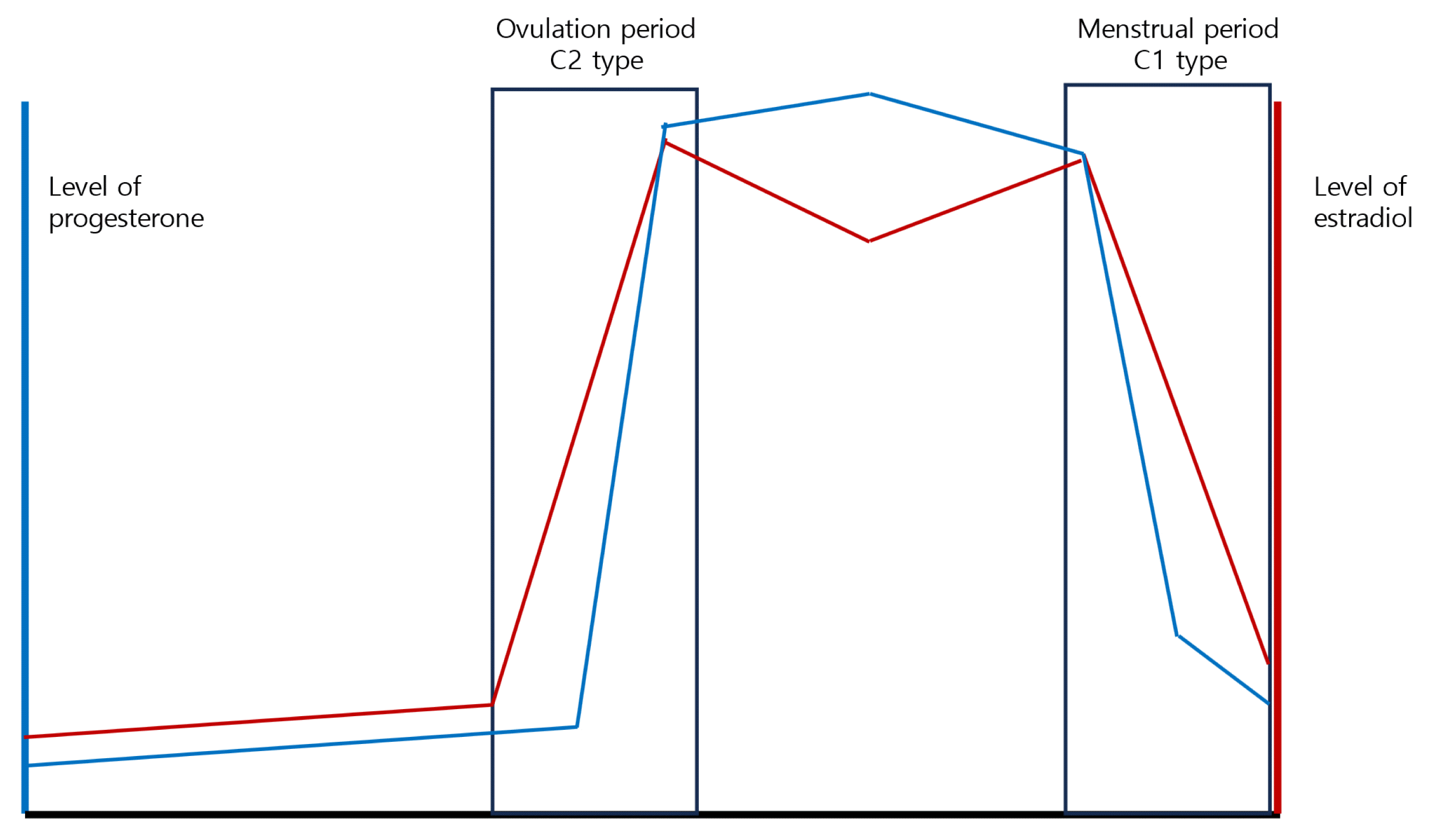
In the context of epilepsy, fluctuating estrogen and progesterone levels throughout the menstrual cycle can influence seizure patterns (Fig. 1). This phenomenon is termed “catamenial epilepsy”.2–4 Estrogen possesses pro-convulsant properties, potentially diminishing the seizure threshold and elevating the likelihood of seizures. Conversely, progesterone, particularly its metabolites like allopregnanolone, exhibits anti-convulsant properties by enhancing the inhibitory function of gamma-aminobutyric acid (GABA), the brain’s primary inhibitory neurotransmitter. Consequently, seizure exacerbation in WWE typically occurs during two specific menstrual cycle phases: firstly, the perimenstrual phase (C1 type), encompassing the days just before and after menstruation’s onset, characterized by a rapid decline in both estrogen and progesterone levels, albeit more pronounced for progesterone; secondly, ovulation (C2 type), marked by elevated estrogen levels and a relative decrease in progesterone levels. In cycles without ovulation, progesterone levels might not rise as expected post ovulation, potentially creating an environment conducive to seizures. If this hormonal pattern consistently repeats in a woman with epilepsy, it could conceivably lead to an overall escalation in seizure frequency (C3 type).

One menstrual cycle and levels of hormones. Rectangular areas represent the period of exacerbation because of high ratio of estrogen to progesterone level. C3 type (not shown here) represents the catamenial epilepsy with anovulatory cycles.
Following menopause, the ovaries cease to produce estrogen and progesterone at the previous levels. This hormonal shift can influence seizure patterns, though the precise relationship remains incompletely understood, as research outcomes have been varied. Certain studies suggest that seizure frequency may rise during the perimenopausal phase, likely due to hormone level fluctuations, before declining in the postmenopausal phase.
Limited options exist for treating catamenial epilepsy, and no therapy has received approval from the United States Food and Drug Administration. A randomized, double-blind, placebo-controlled trial assessed the use of natural progesterone (n=294) for treatment. Statistically significant seizure reduction was only observed for the C1 type.5 Synthetic progestins were explored to achieve complete suppression of the menstrual cycle, resulting in a 39.0% seizure reduction.6 Additionally, clobazam prescribed over a 10-day period during seizure exacerbation demonstrated superiority over a placebo, with confirmed efficacy.7 A trial involving acetazolamide yielded a 40.0% reduction in seizures.8
Fertility Challenges and Epilepsy
Research indicates that WWE may experience lower fertility rates than those without epilepsy. A web-based study conducted in the USA revealed a 9.2% risk of infertility and a 20.7% risk of impaired fecundity among WWE. Impaired fecundity showed a higher trend in ASD polytherapy compared to no ASD use.9 Factors contributing to reduced fertility in WWE are likely multifaceted, involving seizures’ effects, the influence of ASD (ASDs), and conditions like polycystic ovarian syndrome that impact fertility.
WWE also face an elevated risk of spontaneous abortion. The causes encompass both direct seizure effects and the teratogenic impact of specific ASDs. One population-based cohort study demonstrated a higher risk of spontaneous abortion in WWE, with exposure to the ASD valproic acid linked to increased risk.10
Exogenous hormones used in in vitro fertilization (IVF) could potentially exacerbate seizures. Gonadotropins used for ovarian stimulation and exogenous estrogen for menstrual synchronization can elevate systemic estrogen levels. There are case reports of seizure exacerbation during hormonal treatment.11 Nonetheless, the success of reproductive treatments such as IVF appears comparable to that of women without epilepsy.12
Epilepsy and ASDs might impact hormonal treatment during IVF. Specific ASDs can affect steroid hormone metabolism, potentially diminishing the effectiveness of hormone treatments in IVF. Seizures and ASDs can disrupt hormonal cycles, influencing the timing and response to IVF treatments.
The use of ASDs can also influence fertility in both men and women. In women, enzyme-inducing ASDs can lead to hormonal imbalances that disrupt the menstrual cycle and ovulation, consequently reducing fertility.9 For men, studies suggest that long-term use of certain ASDs can impact sexual function, manifesting as decreased libido, erectile dysfunction, and changes in serum testosterone levels.13 Certain ASDs have been associated with adverse effects on sperm count and motility, potentially affecting male fertility.14
Contraception Considerations for WWE
Approximately 50.0% of pregnancies in WWE are unplanned, a rate similar to that observed in the general population.15 The average timeframe for realizing an unplanned pregnancy was 6.5 weeks. According to the Epilepsy Birth Control Registry’s web-based survey, 33.6% of WWE did not use highly effective contraception.16
Various contraception methods are available for WWE. Barrier methods are safe and effective as they remain unaffected by antiepileptic medications. Intrauterine devices (IUDs), encompassing both copper and hormonal options, provide safe and highly effective birth control for WWE, independent of epilepsy medications. Notably, there is a 6.75 times higher risk of increased seizures with hormonal methods compared to epilepsy patients employing barrier methods.17 The intrauterine devices have emerged as the method of choices with the lowest failure rate for WWE.18
For women taking medications metabolized by hepatic cytochrome P450 enzymes, oral contraceptive failure can occur. Hepatic cytochrome P450 systems metabolize female sex steroids, thus impacting oral contraceptive effectiveness.19 ASDs, especially enzyme-inducing ones, can accelerate the metabolism of hormones in oral contraceptives (estrogens and progestogens), diminishing their efficacy. ASDs with this effect include phenytoin, carbamazepine, phenobarbital, primidone, topiramate (at high doses), oxcarbazepine, felbamate, and rufinamide.
Conversely, several ASDs primarily excreted via the kidneys do not induce liver enzymes and hence do not interfere with oral contraceptive pills.20 These include lamotrigine, levetiracetam, gabapentin, pregabalin, ethosuximide, and vigabatrin. However, oral contraceptives can reduce serum levels of lamotrigine, potentially affecting its effectiveness. The hormone estrogen in contraceptive pills accelerates the metabolism of lamotrigine in the liver, leading to decreased overall levels in the body. Therefore, women using both lamotrigine and hormonal contraceptives may encounter increased seizure frequency. Furthermore, during hormone-free intervals (e.g., pill-free weeks), lamotrigine levels could rise, potentially causing side effects. Regular monitoring of lamotrigine levels is advisable when starting or stopping hormonal contraceptives.19,21
For women on enzyme-inducing ASDs, progesterone-only pills or injectables can be used, possibly requiring higher doses of progestogen-only pills.22 The efficacy of injectables like Depo-Provera is not significantly affected by ASDs. While enzyme-inducing ASDs may reduce the effectiveness of contraceptive implants, these can still be employed effectively with non-enzyme-inducing ASDs. Emergency contraception methods using progestogen-only pills (levonorgestrel or ulipristal acetate) might be less effective for women taking enzyme-inducing ASD. In this context, using a copper IUD for emergency contraception remains unaffected by these medications and proves highly effective.
Pharmacokinetic Changes of ASDs during Pregnancy
Pregnancy can induce significant alterations in the pharmacokinetics of ASDs, potentially influencing seizure control and the susceptibility to drug-related adverse effects. These changes predominantly arise from physiological transformations inherent to pregnancy.23–25
Throughout pregnancy, an augmentation in total body water and fat reserves occurs, leading to an increased distribution volume for hydrophilic and lipophilic drugs respectively, which could consequently reduce serum drug concentrations. A considerable number of ASDs undergo hepatic metabolism. Pregnancy can enhance the activity of multiple cytochrome P450 enzymes and uridine diphosphate glucuronosyltransferases (UGTs), crucial players in drug metabolism. Consequently, this heightened enzymatic activity might elevate the clearance of ASDs, thereby diminishing serum drug levels.
Pregnancy is linked to a reduction in serum albumin levels. Given that numerous ASDs exhibit high protein binding, this decrease can augment the free (unbound) drug fraction, the pharmacologically active form. Furthermore, pregnancy coincides with heightened renal blood flow and glomerular filtration rate, potentially accelerating the renal clearance of drugs excreted by the kidneys. Additionally, WWE might be prescribed other medications, introducing the possibility of drug-drug interactions that further complicate management.
Such changes imply that pregnant WWE might necessitate ASD dosage adjustments to uphold seizure control while mitigating potential fetal risks. Regular monitoring of serum drug levels and necessary dosage adjustments are imperative during pregnancy.26–29
Specific ASD Considerations
Levetiracetam
Pregnancy is known to enhance the clearance of levetiracetam, resulting in reduced serum concentrations for most of the pregnancy duration. Accordingly, dose adjustments might be essential to maintain therapeutic levels.
Lamotrigine
The metabolism of lamotrigine is markedly accelerated during pregnancy, chiefly due to heightened UGT enzyme activity, especially UGT1A4. This can lead to a substantial decline in serum concentrations, particularly evident during the third trimester. Close monitoring and dose adaptation are generally necessary. Lamotrigine levels quickly rebound post-delivery, often warranting postpartum dose reduction.
Oxcarbazepine and carbamazepine
Metabolized by cytochrome P450 enzymes, these ASDs experience increased enzymatic activity during pregnancy. This phenomenon may lead to decreased serum concentrations, prompting potential dose modifications. Carbamazepine’s auto-induction of metabolism can further exacerbate this reduction. The most pronounced concentration decrease usually occurs in the third trimester.
Topiramate and zonisamide
Both topiramate and zonisamide are eliminated through renal excretion, and the heightened renal clearance during pregnancy can result in decreased drug levels. Dose adjustments might be necessary.
Phenobarbital and primidone
Pregnancy can enhance the metabolism of phenobarbital and primidone, leading to reduced serum concentrations. The most pronounced decline typically occurs during the first trimester. Adjustments to the dosage might be essential to uphold therapeutic levels.
Valproate
While valproate’s clearance might increase during pregnancy, this change is often less significant compared to other drugs. Nonetheless, individual patients might require dosage adjustments. Serum concentration reduction occurs consistently throughout pregnancy.
Change in Seizure Frequency during Pregnancy
Pregnancy entails various alterations that can impact seizure frequency in WWE. Physiological and hormonal shifts associated with pregnancy can lead to heightened or diminished seizure activity. Comparable to the menstrual cycle, hormonal fluctuations during pregnancy can influence seizure occurrence. Elevated levels of progesterone and estrogen during pregnancy can affect seizure control. Progesterone, usually possessing anticonvulsant properties, is metabolized into allopregnanolone, a neurosteroid that enhances GABAergic inhibition, potentially reducing seizure activity. However, the exact impact of elevated estrogen levels, which can trigger seizures, remains less clear during pregnancy. The intricate interplay between these hormones and their proportions throughout pregnancy may contribute to altered seizure frequency.30
Modifications in drug metabolism during pregnancy can significantly influence seizure management. Pregnancy can accelerate the metabolism and reduce serum levels of ASD, heightening the risk of breakthrough seizures. Consistent monitoring of antiepileptic drug levels and appropriate dose adjustments are crucial in managing this risk.31 Pregnancy, being a potentially stressful period, could also cause sleep disturbances, a recognized seizure trigger.32
As per a study published in Neurology by European Registry of ASD and Pregnancy (EURAP) researchers, out of 3,607 pregnancies, 37.6% experienced an increase in seizure frequency during pregnancy.33 Several factors contribute to this change. Alterations in drug levels during pregnancy can augment the seizure risk. In the same study, lower plasma levels of lamotrigine and oxcarbazepine were linked to a significantly higher risk of worsened seizures. Ceasing or reducing ASD due to concerns about teratogenic effects could potentially raise the seizure risk. Certain epilepsy types might be more susceptible to heightened seizure frequency. For instance, focal epilepsy exhibited a greater risk of increased seizures compared to generalized epilepsy. Women who had a high seizure frequency in the 9 months preceding pregnancy were more prone to experiencing heightened seizures during pregnancy.33
Congenital Malformations in WWE
It is imperative to acknowledge that multiple factors can impact the risk of congenital malformations, including the specific ASD employed, its dosage, the utilization of multiple ASDs, and the mother’s overall health status. While numerous ASDs have been linked to an elevated risk of congenital malformations, it is crucial to balance this risk against the potential hazards of uncontrolled seizures to both the mother and the fetus. Here are specific insights into the potential risks associated with certain ASDs.
Valproate
Among ASDs, the use of valproate in pregnant women is linked to the highest risk of major congenital malformations, encompassing neural tube defects, facial clefts, hypospadias, and heart defects.34
Carbamazepine
Associated with a moderate risk of congenital malformations, carbamazepine’s effects include neural tube defects, craniofacial anomalies, fingernail hypoplasia, and developmental delay.35
Phenobarbital and primidone
These ASDs are correlated with an increased risk of congenital malformations such as cleft lip and palate, heart defects, and growth retardation. Their use during pregnancy is generally limited to cases where alternative treatments prove ineffective.36
Phenytoin
Linked to fetal hydantoin syndrome, phenytoin usage during pregnancy can result in craniofacial abnormalities, nail and digit hypoplasia, growth retardation, and developmental delay.37
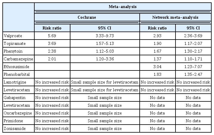
Systematic review and meta-analysis revealed a higher calculated incidence of congenital malformations in WWE compared to healthy women (7.08% vs. 2.28%).38 The incidence was most pronounced for ASD polytherapy (16.78%). Valproate exhibited the highest incidence of congenital malformations, with a rate of 10.73% for valproate monotherapy. Modern ASDs like levetiracetam, gabapentin, oxcarbazepine, topiramate, and zonisamide are increasingly administered to WWE. Assessing the risk of major congenital malformations (MCM) tied to these newer ASDs remains crucial. A Cochrane meta-analysis of 31 observational studies demonstrated that the highest MCM risk ratio compared to pregnancy without epilepsy was observed with valproate, followed by topiramate, phenobarbital, phenytoin, and carbamazepine (Table 1). No heightened risk was identified with lamotrigine. While caution is warranted due to limited sample size, no elevated risk was noted for gabapentin, levetiracetam, oxcarbazepine, primidone, or zonisamide during pregnancy.39
A network analysis disclosed significantly escalated MCM risks for specific ASD monotherapies. Ethosuximide showed the highest risk, followed by valproate, topiramate, phenobarbital, phenytoin, and carbamazepine, compared to controls. No increased risk was associated with lamotrigine or levetiracetam (Table 1).40
Teratogenic Risks and ASDs in Pregnancy Registry
Several global registries, including the EURAP, the North American Antiepileptic Drug Pregnancy Registry, UK and Ireland Epilepsy and Pregnancy Registries, and the Nordic Registry, compile data on pregnant WWE. These registries have significantly contributed to our comprehension of the risks tied to ASD utilization during pregnancy.
A study utilizing data from EURAP indicated diverse teratogenic risks based on different ASDs and dosages (Table 2).41 Monotherapy with valproate exhibited the highest risk of major congenital malformations, followed by phenobarbital and phenytoin. The risks associated with MCM for lamotrigine, levetiracetam, and oxcarbazepine remained within the range reported in the general population. Insights from the North American ASD Pregnancy Registry unveiled a higher risk of major malformations in infants exposed to valproate or phenobarbital compared to newer ASDs like lamotrigine and levetiracetam.42 In utero exposure to topiramate correlated with an elevated risk of cleft lip when contrasted with a reference population. Recent updates from the UK and Ireland Epilepsy and Pregnancy Registries underscored that valproate exposure during pregnancy heightened the risk of major congenital malformations more than lamotrigine and carbamazepine.43 The Nordic Registry showcased an increased dose-dependent MCM risk with valproate and topiramate,44 while no differing MCM risk was noted for carbamazepine, oxcarbazepine, or levetiracetam.
For many ASDs, the risk of congenital malformations is contingent on the dosage, with higher doses linked to greater risks. Hence, during pregnancy, it is generally recommended to utilize the lowest effective dose of ASDs (Table 3).41–44 The employment of multiple ASDs (polytherapy) typically correlates with a higher risk of congenital malformations compared to single ASD use (monotherapy). This could stem from additive or synergistic drug effects.45 However, the presence of either valproate or topiramate in polytherapy might primarily contribute to the heightened malformation rate.46
Balancing Medication Needs and Safety
It is imperative to recognize that refraining from necessary epilepsy medication can be detrimental to both the mother and the baby. Uncontrolled seizures can lead to harm, pregnancy loss, and even maternal mortality. Therefore, pregnant women or those planning pregnancy with epilepsy should collaborate closely with their healthcare providers to effectively manage their condition and medication. Ideally, pregnancy planning and preconception counseling should be conducted for all women with childbearing potential affected by epilepsy. This facilitates the optimization of ASD therapy to diminish seizure and teratogenic risks.47, 48
Understanding the potential congenital malformations associated with ASDs is also essential. Commonly observed malformations encompass neural tube defects, congenital heart diseases, orofacial clefts, intestinal atresia, and urogenital defects.49,50 Valproate is linked to various malformation subtypes, including nervous system, cardiac anomalies, oral clefts, clubfoot, and hypospadias.39,44,50 Barbiturates show a high frequency of cardiac malformations. High doses of topiramate are associated with an elevated risk of cleft lip or palate.51
Shifting Prescription Trends
Over the past two decades, there have been discernible shifts in the prescription patterns of ASDs. In the earlier years, first-generation ASDs like phenytoin, carbamazepine, and valproate were prevalent. Nevertheless, due to their potential for teratogenicity, the capacity to induce birth defects, the medical community progressively veered towards newer second- and third-generation ASDs, such as lamotrigine and levetiracetam.52 This transition corresponded with a noticeable reduction in the prevalence of MCMs over this time period.53
ASD Impact on Offspring IQ
The intricate and evolving relationship between ASD (ASDs) and the cognitive development of offspring born to mothers with epilepsy warrants exploration. Several studies indicate that prenatal exposure to valproate can contribute to lower IQ in children. In a prospective observational multicenter study involving monotherapy in the USA and the UK, children exposed to valproate in utero exhibited notably lower IQ scores at the age of 3 compared to those exposed to alternative ASD.54 Specifically, the mean IQ was measured at 101 for lamotrigine-exposed children, 99 for phenytoin-exposed, 98 for carbamazepine-exposed, and 92 for valproate-exposed.
The Neurodevelopmental Effects of ASD (NEAD) study conducted by Inoyama et al.55 found that children exposed to valproate during gestation displayed significantly diminished IQ scores at the age of 6 in comparison to children exposed to other ASDs. Interestingly, the NEAD study also highlighted that children exposed to carbamazepine, lamotrigine, or phenytoin in utero did not exhibit substantial cognitive disparities when compared to the general population. Nonetheless, the long-term safety profiles of these drugs remain relatively unconfirmed. In certain studies, such as one by Shallcross et al.,56 levetiracetam appeared to potentially entail a lower risk of neurodevelopmental impairment when juxtaposed with valproate.
Upon analysis, the adjusted mean IQ at 6 years was found to be 9.7 points lower for children exposed to high doses of valproate (>800 mg), encompassing verbal, nonverbal, and spatial domains.57 High doses of valproate also exhibited an eightfold increased need for special educational intervention. Conversely, low doses of valproate were not correlated with decreased IQ, yet they did show associations with diminished verbal abilities and a sixfold increase in the requirement for special educational intervention. In contrast, in utero exposure to carbamazepine and lamotrigine did not notably impact IQ, although carbamazepine did seem to be linked with reduced verbal abilities.
In a prospective observational study involving children aged 6 or 7 years, those exposed to valproate demonstrated lower performance across all six neurocognitive domains, particularly in language, in comparison to children exposed to carbamazepine, lamotrigine, or levetiracetam.58
Other Effects on Offspring
In a cohort study, prenatal exposure to substances such as topiramate, valproate, and several duotherapies, including levetiracetam with carbamazepine and lamotrigine with topiramate, were correlated with heightened risks of neurodevelopmental disorders like autism spectrum disorders and intellectual disability.59 Conversely, there was a lack of consistently increased risks for neurodevelopmental disorders following prenatal exposure to monotherapy with lamotrigine, levetiracetam, carbamazepine, oxcarbazepine, gabapentin, pregabalin, clonazepam, or phenobarbital. Notably, topiramate and zonisamide were associated with instances of infants being small for gestational age concerning head circumference as well as birth weight.60
Role of Folate in Pregnant WWE
Folic acid is indispensable for numerous metabolic pathways, particularly in nucleic acid formation, especially during the conversion of homocysteine to methionine. Insufficient folic acid has been linked to orofacial clefts, cardiovascular malformations, urogenital anomalies, and limb irregularities in WWE, a phenomenon more pronounced with the usage of enzyme-inducing ASDs.61
Supplementing with folic acid exceeding 400 mcg/d during the initial stages of pregnancy (first 12 weeks) not only reduces the incidence of major congenital malformations but also holds beneficial effects on IQ and language development.62,63 Furthermore, this supplementation exhibits advantages in mitigating the risks of the development of autistic traits in offspring exposed to lamotrigine, carbamazepine, and valproate.64,65 In WWE using ASDs, the use of periconceptual folic acid has also been associated with a lower risk of preterm birth.66 The American Academy of Neurology practice guidelines recommend a range of 0.4 mg/d to 4 mg/d of preconceptional folic acid supplementation for WWE of childbearing age.
However, an observational cohort study conducted in Nordic countries indicated that high doses of folate (mean 4.3 mg) were linked to an elevated risk of cancer in children of mothers with epilepsy. Although the incidence of cancer is low (1.4% in children exposed to high-dose folate compared to 0.6% in those unexposed), caution is advisable when prescribing high doses of folate.67
Direct Impact of Seizures on Fetus and Delivery
It is challenging to precisely determine the relative impact of various seizure types.68 Two reports noted the effects of focal seizures with impaired consciousness on the fetus, leading to alterations in fetal heart rate during delivery and transient maternal hypoxia.69,70 Fortunately, both cases resulted in the birth of healthy children.
An examination of the effects of seizures on fetuses in the pilocarpine-induced epilepsy model revealed significant repercussions on specific hippocampal interneurons in the offspring of rats experiencing epileptic seizures during pregnancy.71 The same animal model demonstrated that offspring from epileptic mothers exhibited motor coordination deficits.72 Placentas from epileptic rat models displayed areas of ischemic infarction. Additionally, rats exposed to seizures in utero displayed impaired social behavior compared to those with no intrauterine seizure exposure.73
Furthermore, changes in fetal heart rate associated with generalized tonic-clonic seizures (GTCs) were identified. Two cases demonstrated a mixture of tachycardia and bradycardia shortly after GTCs.74 Another case reported fatal intracranial hemorrhage in utero following maternal GTCs.75 It is suggested that experiencing over five GTCs during pregnancy might be linked to detrimental effects on neurodevelopment or lower IQ.76,77
Epilepsy and Other Reproductive Outcomes
A meta-analysis of 38 studies revealed that WWE had higher odds of experiencing various reproductive outcomes compared to those without epilepsy. The odds ratios (OR) for WWE were as follows: spontaneous abortion (OR, 1.54), antepartum hemorrhage (OR, 1.49), postpartum hemorrhage (OR, 1.29), hypertensive disorder (OR, 1.37), induction of labor (OR, 1.67), cesarean section (OR, 1.40), any preterm birth (OR, 1.16), and fetal growth restriction (OR, 1.26).78 In Norway’s compulsory Medical Birth Registry, infants exposed to ASDs were more likely to be associated with preterm delivery, low birth weight, small head circumference, and low Apgar scores when compared to non-epilepsy controls.79 The occurrence of small-for-gestational-age infants was higher in both ASD-exposed and unexposed epilepsy patients. WWE had a significantly elevated risk of death during delivery hospitalization (80 deaths per 100,000 pregnancies) compared to women without epilepsy (6 deaths per 100,000 pregnancies), with an OR of 11.46.80
Breastfeeding
Breastfeeding is generally recommended for most WWE, even those taking ASDs. According to guidelines from the American Academy of Neurology and the International League Against Epilepsy, the potential benefits of breastfeeding often outweigh the potential risks, particularly if the mother’s epilepsy is well-managed on her medications.81
The relative infant dose (RID) serves as a measure of the medication exposure for a breastfeeding infant. It is calculated as the infant’s dose (in mg/kg/day) divided by the mother’s dose (in mg/kg/day), expressed as a percentage. A RID of less than 10.0% is typically deemed safe for breastfeeding, indicating that the infant is exposed to less than 10.0% of the maternal dose adjusted for weight.82 Most ASDs generally have low RIDs, and the safety of breastfeeding while on these medications can depend on various factors, including the specific drug, dosage, timing of doses in relation to feeding times, and the infant’s age. The highest RIDs were as follows: carbamazepine (3.70%), lamotrigine (36.33%), primidone (4.96%), phenobarbital (3.15%), gabapentin (4.37%), valproic acid (1.90%), ethosuximide (31.49%), levetiracetam (12.50%), and topiramate (12.18%).83
While RID is a valuable tool to assess drug safety during breastfeeding, directly measuring infant serum concentrations would be even more informative than breast milk concentrations. Recent studies revealed that most ASD concentrations in the blood samples of breastfed infants were significantly lower than maternal blood concentrations (Table 4).84 Median concentrations of lamotrigine and levetiracetam in infants were 28.9% and 5.3% of maternal plasma levels, respectively. While maternal concentration was a significant factor associated with lamotrigine concentration in infants, it did not have the same effect on levetiracetam concentration in infants. In general, most ASDs are considered relatively safe for breastfeeding.85
Furthermore, evidence suggests that breastfeeding while on ASDs might have a positive impact on the infant’s cognitive development. There was no significant difference in IQ at age 3 between children whose mothers breastfed while on ASDs and those who did not.86,87 Similarly, ongoing studies indicated that breastfeeding had no adverse effects on ASD-exposed infants at age 6 and that breastfed children exhibited higher IQ scores and improved verbal abilities.88
Vitamin K Supplementation
Enzyme-inducing ASD (EIASDs) may potentially lead to hemorrhagic complications in newborns due to increased metabolism of vitamin K. However, a prospective cohort study monitoring 662 WWE using EIASDs found that bleeding complications occurred in 0.7% of newborns born to mothers using EIASDs and 0.4% in control subjects.89 The American Academy of Neurology updated its guidelines in 2009, stating that there was insufficient evidence to establish whether newborns of WWE had a significantly higher risk of hemorrhagic complications.90
Depression and Anxiety
Peripartum depression and anxiety are common complications among women during childbirth, encompassing major and minor depressive episodes and anxiety disorders during pregnancy or the postpartum period.91,92 Individuals with epilepsy are particularly susceptible to developing depression and anxiety.93 A review of current original articles on this topic revealed that the point prevalence of depression ranged from 16.0% to 35.0% in WWE, compared to 9.0% to 12.0% in controls, from the 2nd trimester to 6 months postpartum.94 The highest estimates were observed early in pregnancy and the perinatal period. Factors such as previous psychiatric disease, history of sexual/physical abuse, ASD polytherapy, and high seizure frequency were identified as strong risk factors. Depressed WWE rarely used antidepressant medication during pregnancy. The impact of in utero exposure to ASDs with antidepressants remains unknown, requiring further study. Presently, non-pharmacological treatment or switching to lamotrigine before pregnancy for childbearing women with high risk factors may be considered.
Ideal ASD
An ideal ASD for potential childbearing women and pregnancy should possess the following characteristics: it should not increase the risk of birth defects, ensuring the safety of the fetus at all developmental stages. The drug should not negatively impact the cognitive, emotional, or physical development of the child. It should not affect a woman’s fertility. Ideally, the ASD should be safe for breastfeeding, posing no threat to the infant. The drug should not disrupt hormonal balance, bone health (given the effect of pregnancy on bone density), or other physiological systems in women. Additionally, the drug should not interfere with the absorption or metabolism of essential vitamins like folic acid, crucial for preventing neural tube defects during pregnancy.
Pregnancy induces various physiological changes that influence drug metabolism. An ideal ASD would maintain consistent pharmacokinetics throughout pregnancy. Hormonal and metabolic alterations during pregnancy can affect seizure frequency and severity; therefore, the ASD should remain effective throughout pregnancy. It should not easily traverse the placenta to impact the fetus, and if it does, it should have no adverse effects. As pregnancy might necessitate medication dose adjustments, an ideal drug would have easily manageable and reliable monitoring parameters.
Currently, no ASD possesses all these characteristics. The two most commonly used drugs for WWE are lamotrigine and levetiracetam. The table below offers a comparative analysis of these two medications, based on their ideal characteristics, as outlined in Table 5. A proper understanding of the issues associated with women who have epilepsy is essential for their appropriate treatment. Key points concerning fertility are presented in Table 6.
Notes
The author declare that they have no conflicts of interest.


