A Case of Temporal Onset Partial Seizure Induced by Photic Stimuli
Article information
Abstract
There are only a few case reports of photic stimulation induced partial seizures arising from the temporal lobe. A 12-year-old female with a history of three convulsions was admitted for a diagnostic evaluation. During continuous video-electroencephalogram monitoring, a complex partial seizure with secondary generalization was induced immediately after a photoparoxysmal response with a 15-Hz photic stimulation. This is a rare case of photosensitive temporal lobe seizure.
Introduction
Photic stimulation is a common extrinsic trigger factor in reflex epilepsy. Clinical seizures as well as photoparoxysmal responses can be triggered by an intermittent photic stimulation (IPS), most commonly in generalized epilepsies.1 While some focal seizures are caused by an IPS, most are caused by problems of the occipital lobe or visual cortex.2 Now we are reporting the case of a 12-year-old female patient with a partial seizure triggered by an IPS, proved to be a left temporal lobe seizure, which is supported by continuous video-EEG monitoring.
Case
A 12-year-old girl with a history of three convulsions was hospitalized for diagnostic evaluation including continuous video-EEG monitoring. She began having convulsions 1 year ago. She did not remember her prodromal symptoms or auras. There was no witness during the first two convulsions. During the last convulsion, her mother heard a crashing sound followed by regular gasping and sounds of her striking the floor. However, she did not see the convulsive movement because of the locked door. After the convulsions, the patient was confused for several minutes, and complained of nausea and generalized myalgia. She did not have any history of perinatal problems, developmental delay or febrile convulsions. She did not have any relatives with epilepsy in her family. In a neurological examination, there were no abnormal findings. Her MRI scan with epilepsy protocol was also normal.
During the continuous video-EEG monitoring, the inter-ictal EEG was normal. Only one clinical seizure was detected. Interestingly, this epileptic seizure was induced by photic stimulation. Immediately after a 15-Hz IPS, she went into motionless staring, which lasted about 20 seconds. This was followed by a forced turning of the head to the right and then a secondary generalized tonic-clonic seizure. After a seizure of about 3 minutes, she fell asleep. The EEG during this event (Fig. 1) showed a photoparoxysmal response (generalized spike and waves around 3 Hz during 3 seconds) during the 15-Hz IPS followed by ictal discharge with an onset of rhythmic alpha activity over the left temporal area (T5 maximum). She did not complain or remember any visual problems, such as loss of sight, flickering lights, or visual hallucinations during the seizure or postictal period.
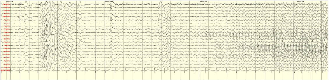
Left temporal lobe seizure induced photic stimulation. Patient's EEG record is showing generalized photo-paroxysmal response during a stimulus of photic stimulation (15-Hz) followed by rhythmic alpha waves over left temporal area (maximum at T5).
The patient was given levetiracetam, 1,000 mg/d and had no seizures for 6 months. The follow-up EEG revealed photoparoxysmal response, however, no seizure was elicited by IPS.
Discussion
Epileptic seizures triggered by photic stimulation are usually associated with idiopathic generalized epilepsies. A quarter of patients with spontaneous seizures and EEG photosensitivity belong to a variety of idiopathic epileptic syndromes, such as juvenile myoclonic epilepsy, childhood absence epilepsy and epilepsy with grand mal on awakening.1 The overall annual incidence of cases with newly presenting seizures and unequivocal photosensitivity in the United Kingdom is 2% of all new cases with epileptic seizures.3 The photoparoxysmal response pattern is usually a generalized polyspikes and slow waves. This wave is highly associated with clinical photosensitivity and clinical events such as jerks or subjective sensations.4
Since there have been several reports of photic stimulation induced occipital lobe seizures, some investigators believe that the origin of focal seizures that are photosensitive can only be occipital.1,5–7 In these cases, corticocortical non-specific visual afferents from the visual area to the frontorolandic cortex seem to be involved in photosensitivity and subsequent spreading occurs through cortical pathways and possibly through thalamocortical pathways.4
There have been a few reports describing photosensitive focal seizure arising from the temporal lobe. Benbadis et al. has proposed that true temporal lobe epilepsy can also be photosensitive.7 Their case is supported by electroclinical data and confirmed by a freedom from seizures after a temporal lobectomy. In 1999, another three cases of complex partial seizures provoked by photic stimulation were reported.8,9 The authors suggested the possibility of activation of distant epileptogenic structure in temporal lobe by photic stimulation of the normal occipital cortex through cortico-cortical transmission. Inoue et al. presented a case diagnosed from clinical symptoms, EEGs, and a single photon emission computer tomography.10 They suggested the photic-driving occipital pathways and afferent pathways from the extraocular muscles or orbicularis oculi can cause temporal lobe seizures.
There may be a possibility of the coexistence of idiopathic generalized epilepsy and focal epilepsy.11–13 Rei Enatus reported a 13-year-old girl with well controlled generalized epilepsy and medically-refractory left temporal epilepsy.14 Wolf and Goosses found that 12 of 103 patients with photic-induced seizures had focal epilepsy. In 8 cases, there was associated evidence of generalized epilepsy and the photic-induced seizures were thought to be generalized. In our case, however, according to medical history, semiology and video-EEG monitoring, there was no evidence of the coexistence of idiopathic generalized epilepsy. The only seizure that we did observe was a complex partial seizure with secondary generalization of left temporal lobe onset. This is a rare case of photic stimulation induced focal seizure from the temporal lobe.