Introduction
Subacute encephalopathy with seizures in chronic alcoholism (SESA) syndrome is a rare clinical manifestation in patients with chronic alcohol abuse. Typical clinical features are characterized by seizures, alteration of mental functions, focal neurological deficit, and focal electroencephalography (EEG) abnormality.1 In most reports, cortical, but not subcortical, lesions are detected.1–3 We herein report the case of a patient with chronic alcoholism who presented with partial nonconvulsive status epilepticus (NCSE) associated with a thalamic lesion that was detected by Magnetic Resonance Imaging (MRI).
Case
A 52-year-old man visited our hospital because of prolonged confusion and abnormal behavior after a generalized tonic-clonic seizure. He had a history of chronic alcohol abuse (approximately 80 g/day). He developed a seizure 38 h after his last drink. He had a previous history of an alcohol withdrawal seizure a month before visiting our hospital. He was irritable, agitated, and violent. His speech was found to be incoherent and he did not obey any verbal command. Automatisms or focal motor seizures were not observed. A routine laboratory test revealed an increase in hepatic enzymes and alkaline phosphatase. Other electrolyte imbalance was not noted. Pleocytosis or protein elevation was not observed on cerebrospinal fluid analysis.
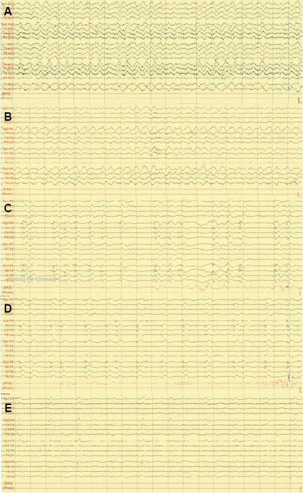
His EEG revealed a continuous partial seizure activity in the hemisphere (Fig. 1A, B). A brain MRI revealed a high-signal-intensity lesion in the right thalamus (Fig. 2A, B). Carbamazepine was administered per orally at a loading dose of 15 mg/kg and a maintenance dose of 300 mg twice a day. On the third day after admission, the patient became free of confusion and his speech improved gradually. Follow-up EEG performed at the third day of admission and the fifth day of admission revealed periodic lateralized epileptiform discharges (PLEDs) in the right hemisphere with gradual reduction of frequency and amplitude (Fig. 1C, D, E). He fully recovered to a premorbid state and was discharged. Abnormal discharge and right thalamic lesion were no longer observed in the follow-up EEG and brain MRI after 6 months (Fig. 2C). Seizures were not observed during the 6-month follow-up.
Discussion
Alcohol withdrawal is the most common cause of seizures in patients with chronic alcoholism, which are usually followed by delirium tremens. Because of the similarity in symptoms, patients with NCSE are often misdiagnosed with delirium tremens or postictal confusion. SESA syndrome usually presents with NCSE, often not associated with apparent symptoms of seizures.2,3 EEG provides an important clue to correct diagnosis. In our patient, there was no laboratory abnormality on serologic test and cerebrospinal fluid analysis to cause the symptoms. He is not typical to SESA syndrome. However, he could be diagnosed with SESA syndrome presenting with NCSE, considering that the PLEDs in EEG and prolonged confusion with speech disturbance with chronic alcoholism.
The pathomechanism of SESA syndrome is not clearly elucidated. Pre-existing cerebral lesions, metabolic disturbance, and alcohol withdrawal may contribute to neuronal hyper-excitability and produce PLEDs, which may lead to clinical focal seizures.4 PLEDs and focal seizures can be observed in patients with metabolic disturbances without structural brain lesions such as non-ketotic hyperglycemia. Focal lesions found in SESA syndrome are considered secondary to focal seizure activity because they are reversible and are similar to those observed in complex partial status epilepticus.2 Patients with SESA syndrome should be treated with anti-epileptic drugs because of high risk of seizure recurrence.3 The question remains whether SESA syndrome is a distinct disease entity or merely a coincidental presentation of partial NCSE in chronic alcoholic patients with underlying epileptogenic foci. Careful clinical follow-up with EEG and neuroimaging may be helpful to solve this question.
In a previous study, thalamic lesions were observed in prolonged partial status epilepticus.5 These lesions are probably due to the functional connectivity between the cortex and the thalamic nuclei and seizure propagation. The initial EEG of our patient shows continuous prolonged rhythmic epileptiform discharge with morphological variability, which is more compatible with NCSE than PLEDs that are triggered by a subcortical lesion.
In conclusion, our patient had an alcohol-related seizure followed by EEG-demonstrated partial NCSE associated with a focal thalamic lesion. Prolonged confusion with focal ictal pattern is compatible with SESA syndrome but the thalamic lesion on MRI is atypical. To our knowledge, this is a rare case report of atypical SESA syndrome characterized by a subcortical thalamic lesion secondary to partial NCSE.6


