Park and Hong: High Frequency Oscillations in Epilepsy: Detection Methods and Considerations in Clinical Application

Abstract

High frequency oscillations (HFOs) is a brain activity observed in electroencephalography (EEG) in frequency ranges between 80–500 Hz. HFOs can be classified into ripples (80–200 Hz) and fast ripples (200–500 Hz) by their distinctive characteristics. Recent studies reported that both ripples and fast fipples can be regarded as a new biomarker of epileptogenesis and ictogenesis. Previous studies verified that HFOs are clinically important both in patients with mesial temporal lobe epilepsy and neocortical epilepsy. Also, in epilepsy surgery, patients with higher resection ratio of brain regions with HFOs showed better outcome than a group with lower resection ratio. For clinical application of HFOs, it is important to delineate HFOs accurately and discriminate them from artifacts. There have been technical improvements in detecting HFOs by developing various detection algorithms. Still, there is a difficult issue on discriminating clinically important HFOs among detected HFOs, where both quantitative and subjective approaches are suggested. This paper is a review on published HFO studies focused on clinical findings and detection techniques of HFOs as well as tips for clinical applications.

Introduction

High frequency oscillations (HFOs) have drawn attention as a new biomarker of epileptogenesis and ictogenesis during last two decades. This brain activity is observed in high frequency ranges between 80 Hz and 500 Hz. From traditional electroencephalography (EEG) monitoring view (generally less than 70 Hz), HFOs could not be observed visually in conventional scope because this is very small and fast oscillating phenomenon. Technical advances in recording system made it possible to acquire signals in improved temporal resolution and to explore on brain activities in higher frequency bands.
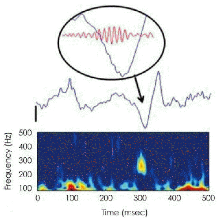
Historically, HFOs of 200 Hz was first observed in hippocampus in normal rat.1 Their clinical importance was highlighted after fast ripples are recorded concentrically on epileptic tissues in mesial temporal lobe epilepsy of rat and humans.2,3 Afterwards HFOs in ripple range are reported to be clinically useful in localizing seizure onset zone (SOZ) in patients with neocortical epilepsy,4 and recordable not only with microelectrodes but also with macroelectrodes.5 Researches on its clinical utility by finding relevance with SOZ and surgical outcomes have been performed.68 Fig. 1 is an example of HFO event from the left parahippocampal gyrus in an epilepsy patient.
HFOs and its related studies have drastically increased in a recent decade. Recent HFO review papers cover different research subjects including recording methods, interactions with other brain rhythms, lesion type difference, influence of sleep cycle, pathological and physiological HFOs, correlation with epileptogenesis and ictogenesis, and its cellular mechanism.911 In this review, we will look through published researches focused on clinical findings and HFO detection techniques with advices to consider in applications.

Clinical findings

Almost thirty percent of epilepsy patients are pharmacoresistent, and epilepsy surgery is an optional treatment for those patients with focal epilepsy.12 Precise delineation and resection of epileptogenic zone is crucial in epilepsy surgery for a outcome of seizure freedom.13 Conventionally intracranial EEG is an essential tool for finding SOZ which is the most important information for the localization of the epileptogenic zone. However, it is not easy to determine an exact epileptogenic zone. Previous resective surgeries based on conventional localization methods did not always ensure postoperative seizure cessation.7,13
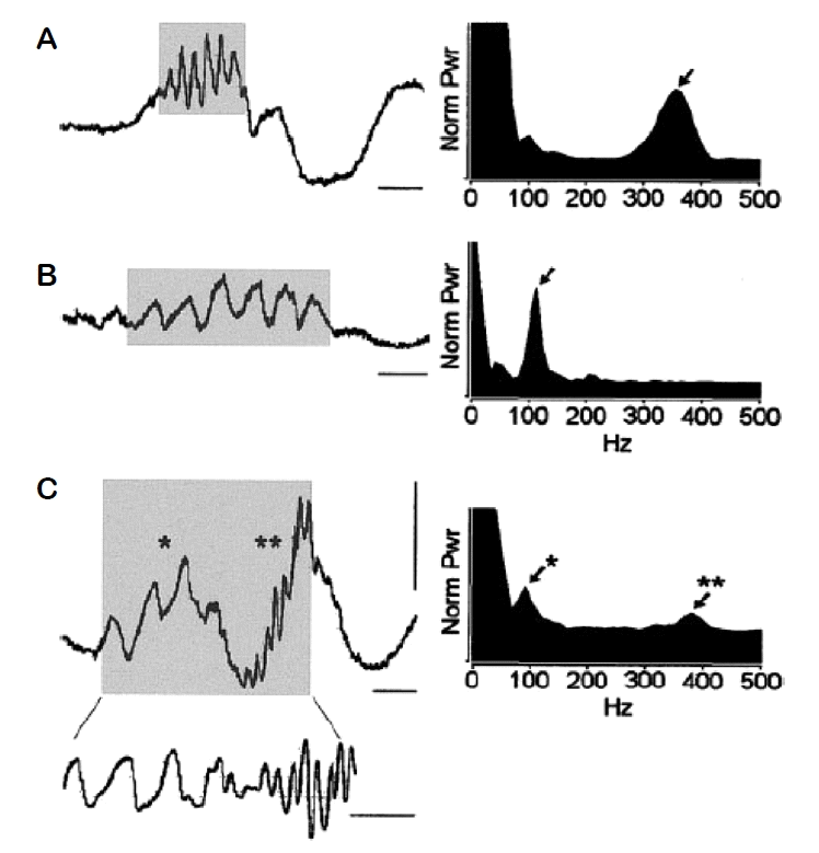
Since HFOs have shown for its possibility as a new biomarker of epileptogenesis in animal studies, human clinical data were started to be investigated in early 2000s. Patients who had a temporal lobe epilepsy and undergone a long term invasive monitoring for a resective surgery were studied in early stage.2,3,1416 Researchers in this time recorded data in hippocampus and mesial temporal lobe structures with microelectrodes. They automatically detected HFOs and quantified data in peak frequency, duration, and amplitudes, etc. HFOs can be classified into two groups of ripples (R; 80–200/250 Hz) and fast ripples (FR; 200/250–500 Hz) by their distinctive characteristics.17 Fig. 2 demonstrates an exemplary morphology of R and FR, and their increased power in a spectral density. Early literatures stated fast ripples appeared briefly with smaller amplitudes than ripples, showing FRs occur exclusively in epileptic tissues. By this observation, researchers at this time speculated its tight correlation of FRs and epileptogenesis. The relation of ripples and epileptogenesis is under a debate and this will be discussed in a later section. A recent study mentioned a peak frequency alone cannot represent the feature of HFO because it can be calculated variously depending on a sampling rate and recorded data.18 Even so, a frequency analysis is critical and it has been utilized in a lot of studies to analyze HFOs.
Neocortical epilepsy is more complex than mesial temporal lobe epilepsy for a surgery because SOZ is localized less clearly and its underlying pathology varies in patients.4,17 The early studies on neocortical epilepsy with subdural macroelectrodes found that HFOs are clinically useful in localizingthe ictal onset zone.4,6 The number of detected HFOs was fewer in neocortical areas compared to the number in mesial temporal areas.6 However, HFOs were detected significantly higher in SOZ than non-SOZ in both mesial temporal lobe epilepsy and neocortical epilepsy.16,1922
HFOs may or may not co-occur with epileptic spikes.6,22 It is reported that HFOs designate SOZ more specifically than interictal spikes.6,23 Contrary to interictal spikes, HFOs do not increase after seizures, but do so after medication reduction, similarly to seizures. This implies that spikes and HFOs have different pathophysiologic mechanisms and that HFOs are more tightly linked to seizures than spikes.24 HFOs and spike show different evolutional phenomenon during interictal to ictal transition. Also, HFOs increased from interictal to preictal periods and spatially restricted; meanwhile, spikes were observed in different regions in interictal, preictal and ictal periods.25
Researches on spasm and semiology found that there are close relations between HFOs and semiology. Fast wave bursts of HFOs and gamma were rapidly spreading when there were epileptic spasms in children.26 In a patient with epileptic spasms, the patient’s HFOs areas were included in resection and had seizure freedom afterwards. The ictal HFOs started before clinical spasm events so that authors suggested that HFOs seem to trigger spasms.27 In patients with Jacksonian seizures, ictal HFOs were confined to focal regions whether conventional EEG spread widely in secondary generalized seizures.28
As the disease severity increased, the higher rates of HFOs were observed.24,29 The relationship of HFOs and seizure activity was found in both intracranial and scalp EEG recordings. The antiepileptic drug reduction study using intracranial EEG suggested a correlation between seizure activity and increase of ripple and fast ripple was found in focal epilepsy.24 In a scalp EEG study, pediatric patients were included: in West syndrome, gamma and ripple range (40–150 Hz) oscillations were reduced after adrenocorticotropic hormone treatment correspondingly as hypsarrhythmia subsided.30 Another study on Rolandic epilepsy visually verified ripples on interictal spikes in children and found that patients having spikes with ripples on them is likely to have more seizures than patients having spikes without ripples, which means the severity of epilepsy syndrome can be checked by ripples on Rolandic spikes.29
Patterns of HFOs were unchanged in different lesion types in a study which included patients with mesial temporal atrophy, focal cortical dysplasia (FCD), and nodular heterotopias.31 However the HFO rates vary with different pathologies. A study found that more HFOs were detected per unit time in FCD, mesial temporal sclerosis, and nodular heterotopias than pathologies with atrophy, polymicrogyria, and tuberous sclerosis.32 Especially in FCD cases, the HFO recording rates were the highest inside the lesion, lower in the peri-leisional and the lowest in remote non-lesional areas. A formerly published study with 22 FCD cases, which focused on comparing HFO rates between FCD type 1 and type 2, found higher HFO rates were observed in type 2 than in type 1. Patients with FCD type 2 had more seizures than those with type 1, so the relationship of epileptic activity and HFO rates could be deduced.33
Regarding surgical outcome studies, patients with higher resection ratio of HFO regions showed better outcome than the group of lower resection ratio.7,17,21,34 In a case report of bilateral mesial temporal epilepsy, the resection of hippocampus in the side with higher HFO recording resulted in seizure cessation for one year.35
Most of postsurgical follow-up studies were based on retrospective perspective, which resection margins were determined based on conventional methods. A Cochrane review by Gloss et al.36 reported two prospective studies which included HFO-generating regions in the determination of resection margin with a postsurgical follow-up over 1 year. Those two studies analyzed ictal HFOs in ripple range recorded on subdural grid, strip and depth electrodes.37,38 The number of included patients was eleven without a control group, where Gloss et al.36 reported an unreliable evidence to draw a conclusion for clinical utility. However, Höller et al.39 supported a clinical significance of HFOs in a meta-analysis of 11 studies focusing on the relationship of HFOs and surgical outcome.

Detection of HFOs

Digital EEG recording systems enabled to transform EEG raw data into a discrete format. From this fact, it is important to consider Nyquist rate in determining recordable maximum frequency range in HFOs. According to Nyquist theorem, a sufficient sampling rate is more than twice of maximum upper bound of interested frequency band. This is for protecting original signal from distortion by an aliasing effect.40 Preferably this value requires being at least three times higher than the maximum frequency of HFOs for a clinical data analysis. The maximum recordable frequency band can be determined depending on the equipment specification. The frequency band range and sampling rate range differ in each study group since EEG recordings were undergone with different equipments with different specifications. Researchers usually set the frequency range based on their research interest, for example, 2 kHz sampling rate for HFO studies of the frequency range of 80–500 Hz.6,8,17,21,23,32,41
The first step to detect HFOs is filtering the signal of high frequency range from raw data by applying a digital band pass filter. This can be set by using a software function unit providing a high-pass filter. The finite impulse response (FIR) filter and the infinite impulse response (IIR) filter are widely used in HFOs researches. FIR is the mostly adopted filter for HFO researches6,19,4248 and its main advantages are free from phase distortion, intrinsically stable, and simply designable.40 The IIR filter is also used in a few researches.49,50 It has its advantage of sharp cut-off and narrow transition width,50 though it is uneasy to make a linear phase40 and have a tendency of oscillating more after filtering compared to the FIR filter.51 In a research with IIR filter, a forward-backward filtering technique was used to prevent a phase distortion.52 The Butterworth filter is also used in studies of ictal high frequency activities or scalp HFOs.4,28
After a band-pass filtering of raw signals, data containing only high frequency components where low frequency components are eliminated would remain. There are two ictal studies using only high pass filter which sufficiently showed a clinical significance of HFOs.35,53 However, in many researches, especially interictal studies, further steps were applied to differentiate HFOs from artifacts and sharp waves. The criteria labeling HFOs, in general, is to select data epochs where at least three oscillations are outstanding from the background.
The detection methods could be manual, automatic, a combination of manual and automatic. First, a manual detection was performed by visually watching a computer monitor screen and by enlarging the time scale of band passed data. This method has been used in Montreal Neurology Institute (MNI) group and their collaborators and other researchers studying ictal HFOs.6,19,25,41,5456 They expanded the time scale of monitor screen as large as 250–530 mm/s or 0.8–7.5 s/page.19,25,45,54 The standard criteria for visual labeling of HFOs was more than four oscillations of which amplitudes stand out from the background in interictal periods, and rhythmic activities continuing at least 300 msec in ictal periods.55,56 The visual analysis has several advantages. It is useful in detecting HFOs with a baseline shift which were recorded especially in deep brain regions and is stable from HFO rate changes affected by a spike rate variability.31 However, a drawback of visual analysis is time-consuming.24 Most of studies with a visual analysis included data with limited time lengths: 3–15 minutes.25,32,33,41 Furthermore, the analysis could be subjective depending on evaluators which might lower the reliability of a study. To make up for this problem, two reviewers marked events independently and computed Cohen’s kappa coefficient to determine whether to accept the events.23,25,32,33,41,57 Coefficient threshold over 0.5 was chosen for a moderate agreement between reviewers23,57 in these studies. In case of the coefficient below the threshold, reviewers reanalyzed the event and reached on a consensus on the marking. This coefficient is recently suggested to be set 0.8 or above to avoid subjective variability and to be acceptable in medical applications.58
Second, an automatic method used for HFO detection has been developed by a software detection program. Mostly those are in-house codes and were made by each research group. Representatively, line length (LL) detector,17,59 root mean square (RMS) detectors15,47 Hilbert detector,60 MNI detector61,62 have been utilized by many research groups. LL, RMS, and Hilbert detectors calculate an energy threshold on the time segment with the assumption that HFOs are rare events.63 On the contrary, MNI detector computes baseline segments earlier then a detected local energy threshold.62 A study compared those detectors displaying different detection results in which some results are concordant and some are not. Each detector designated slightly different event indication and duration.62 This difference might be from the fact that HFO detectors were differently designed. Each institution has different types of electrodes and recording hardware, and different definitions on HFO in amplitudes, durations and etc. Usually detector designers find parameters best fitting with their own data characteristics. Regardless a method to be applied, it would be more important clinically to designate brain areas where HFOs are concentrated.
The advantage of an automatic detection method is that it is time-saving and adjustable in large size data. One thing to be reminded in applying an automatic approach is that some of detected results can include false HFOs affected by artifacts. Fig. 3 illustrates three types of false HFO detection by artifacts. False positive events have different morphological and spectral features from those of true HFOs seen in the Fig. 1.
Representatively, a digital filter characteristic itself can affect the occurrence of false positive events especially when a narrow band filter is applied, which is also known as Gibbs’ phenomena64 or ringing effect. A sharp wave (including epileptic spike) could be detected since its intrinsic attribute of generating ripple-like oscillations after filtering. This filtering effect has been reported in several papers.51,60,65 Even well-designed high pass filtering can include false positive events induced by artifacts. Therefore, post-processing steps may be needed to reject those false HFOs. Either manual (including semi-automated) inspection by human expertise17,34,42,50,52,66,67 or automatic post-processing techniques65,68 were applied to solve this issue.
For manual inspection, a time-frequency (TF) plot is generally used. It decomposes high frequency oscillations into time and frequency domains to enable a mapping of one-dimensional time domain signal into a two-dimensional time–frequency domain representation.69 This method has been widely used in EEG researches and former gamma band studies.
TF plots can show power or energy of HFOs in a form of distinguished blub just as Figs. 1, 2. There is no gold standard in implementing a method of TF plot. It varies depending on computation approaches in representing high resolution of time and frequency components. According to the theory, it is unattainable to get time and frequency component in high resolution all together. The increasing resolution of time will decrease resolution of frequency part, and the same phenomenon occurs in opposite way.70 Previously published papers implemented Morlet wavelet,8,60,71 Morse wavelet,66,72 Stockwell transform,50,61,73 or matching pursuit algorithm.74
This TF plot inspection process of verifying blobs traditionally required tedious human workload. However, this can be partly reduced by supervised classification technique. It trains detector using part of dataset HFOs were labeled manually. The test dataset is applied to trained detector to automatically separate true HFOs from false HFOs. Several researches developed TF plot classification techniques using machine learning technique such as support vector machine (SVM).66
Automatic solutions for finding topographic character of TF plot were developed and applied by several groups.50,68 A research suggested wavelet convolution technique to distinguish HFOs ripples from false HFOs generated by sharp waves ringing effect, and where verification was performed by visual inspection.68 Another research by Burnos et al.50 parameterized transitory power spectrum from TF domain and used it to optimize their HFO detector. They optimized parameters using data of from a patient with mesial temporal lobe epilepsy and tested with five patients of different etiology, which stated to have result of reasonable accuracy.50 The following study by Fedele et al.61 performed on large dataset. They trained their detector parameters for TF domain with 14 patients, and tested it against 54 dataset. The data was intraoperative electrocorticography (ECoG) recordings which HFO events were previously marked by human visual inspection. This detector is the fully automated unsupervised detector and has shown a possibility to substitute human experts as it displayed parallel performance. Those detectors were optimized for data recorded from one medical center, so further variety in dataset will ensure the reliability.
Meanwhile, in a research, automatically detected HFOs (including ripples by sharp wave ringing effect) and resection of the detected region sufficiently showed favorable outcome.75 Considering sharp waves are mostly epileptic spikes indicating epileptogenicity, the authors claimed it is might be needless to sort out false HFOs (just as inspecting TF plot) in clinical use.
The other method to differentiate false HFOs from true HFOs is to extract features from recorded signals. Machine learning studies extracted features of each candidate HFO events so that quantified information of frequency contents and waveform morphology could be analyzed. Both supervised67 and unsupervised65,76 classification underwent in machine learning approach.65,67,76 In studies using artificial neural network, features of LL of amplitude, power and frequency were extracted from filtered signals.42,57
The limitation of the automatic detectors studies is that these developed detectors were optimized for small dataset with limited diversity in epilepsy syndromes. Blanco et al.76 group applied their detector against nine neocortical epilepsy patients and two control patients. Burnos et al.50 optimized parameters using one patient’s data and tested with five patients. The study by Fedele et al.61 performed on large dataset that trained their detector parameters in TF domain from 14 patients, and applied to 54 dataset (including patients in training session with different dataset). This detector showed possibility to substitute human expert by displaying parallel performance. Likewise, the neural network study group applied three patients for training and eight patients data were used for detector evaluation.
It is recommendable to apply large dataset in the future researches because small dataset might cause overfitting problem, which is optimized for certain feature of given data and might to fail to predict future detection reliably. It would be meaningful for following studies to apply fully automated detector to diverse dataset characters (recording methods, recording devices, patient etiologies and etc.) with different hospitals to make a detector with general performance.
Although postsurgical outcomes results were not presented in studies above and performance are needs to be verified by human inspectors, automatic detectors are worth to be implemented. Höller et al.,39 mentioned in their meta-analysis that automatic detectors reasonably perform as good as visual detection.

HFOs for clinical use

It is essential to calculate HFOs for each patient because inter-individual variability of HFOs presents, and the HFOs features also vary enormously between pre- and post-ictal period in different patients. Statistics-based patient-oriented research is the way further researches should go forward.
However, their use in presurgical evaluation will require a quantification in an individual patient due to variability in the properties of HFOs, and differences in spatial sampling and recording electrodes among different patients. While this methodology still needs to be developed, it is likely that any patient-based quantitative approach will need to consider the type of HFO, and possibly anatomical location, and use a statistically-derived threshold to identify significant HFO-generating sites. One retrospective study (Akiyama 2011) involving surgical pediatric patients used an approach that calculated rates of ripple- and fast ripple-frequency HFOs in combination with histogram and bootstrapping analysis to define a threshold to identify high-rate HFO sites.64 In addition to an important result that found more complete resection of high-rate fast ripple-frequency HFO sites was associated with better surgical outcome, the novel patient-oriented HFO quantification used in this study could be appropriate for prospective studies of HFOs in presurgical evaluation.
Then how to select clinically significant HFO sites? Do we need to consider all channels with HFOs although even only small amount of HFO were observed in some channels? Or do we need to count channels which HFO occurrence rate is above a certain level? Previous studies reported that resection of HFO channels above a predefined threshold showed better surgery outcome. In order to delineate putative EZ precisely with the detected HFO, researchers have chosen either quantitative or subjective way to solve this issue. Quantitative methods can be divided into two approaches in general.
One way is to sort channels according to HFO rates and select only high-rate HFO channels based on a predefined threshold. Studies investigated in an interictal period adapted this method. Table 1 summarizes published research papers adapted thresholds with high rate HFO calculation in correlation with surgical outcome. Akiyama et al. and Okanishi et al. used Kittler’s method77 to separate high and low rate channels, afterwards, bootstrapping was applied to make up small sample size.34,78 Cho et al.17 calculated thresholds from the statistical solution with Turkey’s upper fence to select high rate HFOs sorted in maximal ordering. All of the studies above calculated it for R and FR respectively, except a recent study by Fedele et al.73 which defined threshold for channels with the highest rate of R co-occurring with FR. They set the threshold for high rate HFO surpassed 95% of the rate distribution.73 A previous study by the same author on intraoperative ECoG determined a HFO rate threshold of 1 event/min, from clinical basis, describing it as a threshold that best predicting for surgical outcome.61 In all above studies, intracranial EEG was recorded with subdural macroelectrodes and selected analyzing data from non-rapid eye movement (NREM) sleep. Whole duration of analyzed data was 20–30 minutes per patient, except one research studied on 1 minute intraoperative ECoG recording.61 All channels were treated independently for the rate computation. Seizure free was the standard of defining favorable outcome after surgery except one study analyzed correlation between HFO rates and outcome in regression manner.28
There is no clear evidence yet which method is the best solution for determining a precise surgical margin. Burnos et al.50 applied a half maximum threshold, where they also tried Kittler’s method to their data and identified both methods had similar sensitivity and specificity. Eventhough a gold standard in the study was SOZ, not a surgical outcome, it suggests different thresholding methods can result in a similar output.
Another way of quantitative delineation of HFOs channels is to calculate increased HFO element (amplitude or power) values by comparison with predefined baselines. The channels with HFO events exceeding a threshold value were regarded as qualified HFOs channels. This approach was adapted in studies which investigated period of interictal to ictal transition or in brain stimulation as summarized in Table 2. Akiyama et al.28 calculated amplitude in each independent channel to find channels more than 50% of amplitudes increased at the onset part of ictal period compared to interical baseline in each R and FR band. Khosravani et al.44 also computed the relative power ratio between preictal (baseline) and postictal where negative ratio value means significant preictal HFO increase. Those two studies calculated a baseline from interictal or peri-ictal period in channel-wise. Other studies used different approaches other than a channel-wise estimation: Modur et al.79 and Leung et al.80 selected only the channels contained power above the median power of all channels. Modur et al.79 analyzed ictal HFO from the immediate before ictal onset point to few time after electrical seizure, and Leung et al.80 analyzed electrical brain stimulation session where they examined increased discharges or seizures after stimulation.
All studies in Table 2 concluded a resection of designated HFO followed by favorable outcome, where two studies included disabling seizure status in favorable outcome. Similar to the studies above, van ’t Klooster et al.71 used event-related spectral perturbation (ERSP) method to analyze increased power after single pulse brain stimulation. The events representing significant power increase after subtracting the baseline in TF plot were counted as effective one, and they found fast ripples have correlation with resection margin. Even partly engaged in manual process in inspecting TF plot, they quantitatively computed baseline from bootstrapping method by averaging power of the randomly selected epochs among pre-stimulation period.
Quantitative thresholding techniques on HFOs seem to be efficient to determine epileptogenic zone. Each study included patients in different number (2–54) with different pathologies as described in Tables 1, 2. One study collected patients with identical pathology (tuberous sclerosis complex), whether others included various pathologies to broadly analyze the correlation of thresholded HFO region and surgical outcome. Even it was reported that HFOs represent EZ regardless of underlying pathologies,7 later published study claimed that HFO rates are detected in different quantity by different lesion type. Rates were significantly higher in FCD, mesial temporal sclerosis, and nodular heterotopia than in atrophy, polymicrogyria, and tuberous sclerosis.32 Therefore, researchers need to pay attention to apply a thresholding method. In case of a patient who has mixture of two or more pathologies, only the lesion site with a lot of HFOs detected can be ranked in high rate HFOs.
Data of all studies above was recorded with subdural grid, strip and depth electrodes, except two studies by Akiyama et al.28,34 inserted only grid electrodes. Using mixture of grids and depth electrodes in recording EEG might affect in counting HFO rate. As a previous review literature mentioned, different electrode sizes influence recording properties and subdural grids tend to be vulnerable to noise signals than depth electrodes.11 There could be possibility that channels with depth electrode capture more HFOs and result in high rate HFOs.
All literatures above tried to sort out HFO regions clinically significant, but only one study among them reflected the thresholding to epilepsy surgery. The study by Modur et al.79 selected a putative surgical region from continuously evolving ictal HFO channels above median power of all channels, during the first 2 second of ictal onset and the region of 1 cm around SOZ. The more prospective studies would give us an exact view on clinical use of HFOs.38

Unsolved Issues and Current Limitations

One of the unsolved issues is to differentiate physiologic and pathologic HFOs. Early findings reported that ripples tend to represent physiological phenomenon, whereas fast ripples represent pathologic event so regarded more as an epileptic marker.2,14,15 Ripple range has been reported to be associated with memory consolidation1,81 and observed in primary motor cortex.82 However, pathologic ripples also reported to be generated in neocortical cortex,6 and several researches revealed that resected brain areas with ripple have correlation with favorable outcome.17,78 Studies reported that fast ripples can accompany cognitive processes and are spontaneously generated by eloquent brain areas in humans.74,8385 Therefore, we can conclude that ripples and fast ripples may display both physiologic and pathologic aspects.
Some researchers differentiated pathologic HFOs from physiologic HFOs by a wave morphology. One study visually verified waveform and suggested that high rate ripples and fast ripples on flat background activity and its resection was correlated with seizure freedom, whereas high rate ripples in constantly oscillating background was not significantly related to surgical outcome.86 Another study automatically classified HFOs using SVM, and they found pathologic HFOs tend to have higher spectral amplitudes, longer mean durations, and lower mean frequency than physiologic HFOs.59
There is a literature investigated sleep cycle to characterize two types of HFOs. The pathological HFO rates decrease during NREM sleep, whether the physiological HFO rates increase during REM sleep.87 The authors concluded that analysis on the first sleep cycle is the best for HFO study. It is important to label pathologic HFOs yet the clinical application and decision should be performed discreetly.
Currently there are more than hundred HFO research articles published but there is lack of consistency in definitions, recording device, electrodes types, detection methods, patients’ clinical backgrounds. For instance, the frequency ranges subtly differ to research groups: boundary between R and FR sometimes set to 200 or 250 Hz, lower R bound set from 60 to100 Hz, and upper FR range stretch to 600 Hz or more. Therefore, it is required to clarify the recording specifications and detection process and criteria labeling HFOs, and patient’s clinical details for future research outputs. Recent meta-analysis document suggested a guideline for future publications,39 and it is recommended for researchers to reference it for future publication.
Future works can include developing fully automated software to reliably detect pathological HFOs. This will be realizable based on dataset sharing between research institutions, develop effective algorithms, and find adaptive parameters by training and testing with different data properties and various patient information.

Acknowledgements

This research was supported by a grant of the Korea Health Technology R&D Project through the Korea Health Industry Development Institute (KHIDI), funded by the Ministry of Health & Welfare, Republic of Korea (grant number: HI16C1643).

Figure 1
A representative example of high frequency oscillations (HFOs) recorded in intracranial electrocorticography in a patient with neocortical epilepsy. Unfiltered raw trace is displayed as a blue line, and the lower display is its time-frequency decomposition plot. Electroencephalography segment of interest is magnified in a black circle to show HFOs, where red traces represent the signal band-passed for fast ripple (200 to 500 Hz). The black vertical line at the left side of the raw signal corresponds to 200 μV. Fast ripples, brief oscillatory activity (−15 ms) riding on a sharp wave is visible with a spectral peak at around 260 Hz. Modified from reference 8 with permission.
er-9-1-1f1.gif
Figure 2
High frequency oscillations (HFOs) and power spectral analysis. Left: wideband electroencephalography (EEG) traces and corresponding power spectral density histograms (right) illustrate three examples of HFOs recorded in hippocampal (Hip) and entorhinal cortical (EC) areas. Gray part: the segments of wideband EEG that represent HFOs were detected using criteria described in methods and they were used in the power spectral analysis. (A) HFO recorded from a patient within Hip ipsilateral to the side of seizure onset. Power spectral analysis reveals peak spectral frequency at 350 Hz (arrow). (B) Lower-frequency HFO peaked at 110 Hz (arrow) recorded within Hip of another patient was contralateral to the side of seizure onset. (C) HFO recorded from EC contralateral to the seizure onset of the same patient in A. Note how the first 30 ms of the HFO begins as a low frequency oscillation (*90 Hz) that changes to a much higher frequency oscillation (**370 Hz). Gray part: the segment of EEG was band-pass filtered (80–500 Hz) and the gain was increased two times for the clarity of illustration. Calibration bars 0.5 mV for all panels and 5, 10, and 15 ms for (A–C), respectively. Modified from reference 15 with permission.
er-9-1-1f2.gif
Figure 3
Three types of artifacts are shown. The upper most section is raw data, middle is the time-frequency plot of the same epoch above, and the lower most section is power band ratio plot. (A) Type 1: high-frequency transients due to high-pass filtering of sharp components of interictal epileptiform discharges, i.e., discharges with no visible superimposed fast oscillations in the unfiltered signal and/or with absence of isolated fast activity (“blobs”) in time-frequency decomposition plane. (B) Type 2: harmonics of low-frequency, non-sinusoidal signals. (C) Type 3: transient events with amplitudes larger than the global background but not significantly different from the local activity. Modified from reference 8 with permission.
er-9-1-1f3.gif
Table 1
Summary of quantitative thresholding methods for prediction of clinically significant HFO channels: finding high rate HFOs
Study HFO thresholding method Selected threshold value Data epoch Analyzed time period Analyzed data length HFO band range (Hz) Patients Pathologies F/U after resective surgery Standard for favorable outcome
Akiyama et al.28 (2011) Kittler’s method + bootstrap method Channels with HFO rate ≥ 1/min IIC NREM sleep data > 1 hour apart from seizures 10 epochs × 2 minutes R 80–200
FR 200–300*
28 Gliosis, FCD, HS, TSC, microdysgenesis, polymicrogyria > 2 years SZ free
Okanishi et al.78 (2014) Kittler’s method + bootstrap method Channels with HFO rate ≥ 1/min IIC NREM sleep data > 1 hour apart from seizures 10 epochs × 2 minutes R 80–200*
FR >200*
10 TSC > 2 years (mean 58 months) n/a
Cho et al.17 (2014) Turkey’s upper fence Channels with HFO counts ≥ each channels’ threshold IIC SWS data > 4 hours apart from seizures 3 epochs × 10 minutes R 80–200*
FR 200–500*
15 Cortical dyslamination, cavernous angioma, gliosis, HS, FCD, astrocytoma, DNET Mean 26.3 ± 4.8 months SZ free
Fedele et al.61 (2016) Clinical threshold empirical acquired Channels with HFO rate > 1/min IIC Intraoperative recording data 1 minutes R 80–250
FR 250–500*
54 FCD, ODG, MTS, cavernoma > 1 year SZ free
Fedele et al.73 (2017) Standard deviation Channels with HFO rate distribution > 95% IIC SWS data > 3 hours apart from seizures 6 epochs × 5 minutes R 80–250
FR 250–500*
20 MTS, ganglioglioma, FCD, DNET, TSC, gliosis > 1 year SZ free

HFO, high frequency oscillations; F/U, follow up; IIC, interictal; R, ripple; FR, fast ripple; FCD, focal cortical dysplasia; HS, hippocampal sclerosis; TSC, tuberous sclerosis complex; SZ, seizure; NREM, non-rapid eye movement; n/a, not applicable; SWS, slow wave sleep; DNET, dysembryoplastic neuroepithelial tumor; ODG, oligodendroglioma; MTS, mesial temporal sclerosis.

* The R or FR with asterisk indicates the frequency range significantly related to surgical outcome.

In this study, the correlation of the resection ratio of high rate HFO regions and the seizure outcome was analyzed in regression manner.

The patient dataset was acquired by van Klooster et al.88 (2015).

Table 2
Summary of quantitative thresholding methods for prediction of clinically significant HFO channels: calculating baseline
Study HFO thresholding method and selected value Data epoch Analyzed time period Analyzed data length Baseline Baseline data length HFO band range (Hz) Patients Pathologies F/U after resective surgery Standard for favorable outcome
Khosravani et al.44 (2009) Channels relative HFO power ratio (R) > 0 for postictal, R < 0 for preictal HFO IC First 5 seconds of electrical ictal onset 5 seconds Preictal period of 5 seconds before electrical ictal onset (channel-wise) 5 seconds R 100–200*
FR 200–500*
7 HS, DNET, gliosis 8–16 months SZ free
Akiyama et al.28 (2011) HFO channels with amplitude score (calculated from Turkey’s upper fence) ≥ 1.5 IC From the time onset of amplitude score above 0.5 to the ictal period Differ by patients IIC epoch aparted > 10 minutes before the ictal onset (channel-wise) 1 minute R 80–200*
FR 200–300*
2 FCD, gliosis > 2 years SZ free
Modur et al.79 (2011) Channels above median power of all the channels with HFO IC First 2 seconds of electrical ictal onset 2 seconds Power of all channels 2 seconds R ≥ 70* 14 Gliosis, focal dysgenesis Mean 27 months (20–38 months) Engel class I, II
Leung et al.80 (2015) Channels of normalized wavelet power spectrum density above the median power of all channels with HFO Brain stimulation After brain stimulation period < 5 seconds Power of all channels < 5 seconds R ≥ 70* 12 MTS, FCD, cavernous haemangioma, gliosis > 1 year Engel class I, II

HFO, high frequency oscillations; F/U, follow up; R, ripple; IC, ictal; FR, fast ripple; HS, hippocampal sclerosis; DNET, dysembryoplastic neuroepithelial tumor; SZ, seizure; IIC, interictal; FCD, focal cortical dysplasia; MTS, mesial temporal sclerosis.

* The R or FR with asterisk indicates the frequency range significantly related to surgical outcome

References

1. Buzsáki G, Horváth Z, Urioste R, Hetke J, Wise K. High-frequency network oscillation in the hippocampus. Science. 1992;256:1025–7.

2. Bragin A, Engel J, Wilson CL, Fried I, Mathern GW. Hippocampal and entorhinal cortex high-frequency oscillations (100--500 Hz) in human epileptic brain and in kainic acid--treated rats with chronic seizures. Epilepsia. 1999;40:127–37.

3. Bragin A, Engel J, Wilson CL, Fried I, Buzsáki G. High-frequency oscillations in human brain. Hippocampus. 1999;9:137–42.

4. Worrell GA, Parish L, Cranstoun SD, Jonas R, Baltuch G, Litt B. High-frequency oscillations and seizure generation in neocortical epilepsy. Brain. 2004;127:Pt 7. 1496–506.

5. Worrell GA, Gardner AB, Stead SM, et al. High-frequency oscillations in human temporal lobe: simultaneous microwire and clinical macroelectrode recordings. Brain. 2008;131:Pt 4. 928–37.

6. Jacobs J, LeVan P, Chander R, Hall J, Dubeau F, Gotman J. Interictal high-frequency oscillations (80–500 Hz) are an indicator of seizure onset areas independent of spikes in the human epileptic brain. Epilepsia. 2008;49:1893–90.

7. Jacobs J, Zijlmans M, Zelmann R, et al. High-frequency electroencephalographic oscillations correlate with outcome of epilepsy surgery. Ann Neurol. 2010;67:209–20.

8. Cho JR, Joo EY, Koo DL, Hong SC, Hong SB. Clinical utility of interictal high-frequency oscillations recorded with subdural macroelectrodes in partial epilepsy. J Clin Neurol. 2012;8:22–34.

9. Frauscher B, Bartolomei F, Kobayashi K, et al. High-frequency oscillations: the state of clinical research. Epilepsia. 2017;58:1316–29.

10. Jiruska P, Alvarado-Rojas C, Schevon CA, et al. Update on the mechanisms and roles of high-frequency oscillations in seizures and epileptic disorders. Epilepsia. 2017;58:1330–9.

11. Zijlmans M, Worrell GA, Dümpelmann M, et al. How to record high-frequency oscillations in epilepsy: a practical guideline. Epilepsia. 2017;58:1305–15.

12. Schuele SU, Lüders HO. Intractable epilepsy: management and therapeutic alternatives. Lancet Neurol. 2008;7:514–24.

13. Rosenow F, Lüders H. Presurgical evaluation of epilepsy. Brain. 2001;124:Pt 9. 1683–700.

14. Bragin A, Wilson CL, Staba RJ, Reddick M, Fried I, Engel J. Interictal high-frequency oscillations (80–500 Hz) in the human epileptic brain: entorhinal cortex. Ann Neurol. 2002;52:407–15.

15. Staba RJ, Wilson CL, Bragin A, Fried I, Engel J. Quantitative analysis of high-frequency oscillations (80–500 Hz) recorded in human epileptic hippocampus and entorhinal cortex. J Neurophysiol. 2002;88:1743–52.

16. Staba RJ, Wilson CL, Bragin A, Jhung D, Fried I, Engel J. High-frequency oscillations recorded in human medial temporal lobe during sleep. Ann Neurol. 2004;56:108–15.

17. Cho JR, Koo DL, Joo EY, et al. Resection of individually identified high-rate high-frequency oscillations region is associated with favorable outcome in neocortical epilepsy. Epilepsia. 2014;55:1872–83.

18. Gliske SV, Irwin ZT, Chestek C, Stacey WC. Effect of sampling rate and filter settings on High Frequency Oscillation detections. Clin Neurophysiol. 2016;127:3042–50.

19. Jirsch JD, Urrestarazu E, LeVan P, Olivier A, Dubeau F, Gotman J. High-frequency oscillations during human focal seizures. Brain. 2006;129:Pt 6. 1593–608.

20. Malinowska U, Bergey GK, Harezlak J, Jouny CC. Identification of seizure onset zone and preictal state based on characteristics of high frequency oscillations. Clin Neurophysiol. 2015;126:1505–13.

21. Urrestarazu E, Chander R, Dubeau F, Gotman J. Interictal high-frequency oscillations (100–500 Hz) in the intracerebral EEG of epileptic patients. Brain. 2007;130:Pt 9. 2354–66.

22. Urrestarazu E, Jirsch JD, LeVan P, et al. High-frequency intracerebral EEG activity (100–500 Hz) following interictal spikes. Epilepsia. 2006;47:1465–76.

23. Zelmann R, Zijlmans M, Jacobs J, Châtillon CE, Gotman J. Improving the identification of High Frequency Oscillations. Clin Neurophysiol. 2009;120:1457–64.

24. Zijlmans M, Jacobs J, Zelmann R, Dubeau F, Gotman J. High-frequency oscillations mirror disease activity in patients with epilepsy. Neurology. 2009;72:979–86.

25. Zijlmans M, Jacobs J, Kahn YU, Zelmann R, Dubeau F, Gotman J. Ictal and interictal high frequency oscillations in patients with focal epilepsy. Clin Neurophysiol. 2011;122:664–71.

26. Asano E, Juhász C, Shah A, et al. Origin and propagation of epileptic spasms delineated on electrocorticography. Epilepsia. 2005;46:1086–97.

27. Akiyama T, Otsubo H, Ochi A, et al. Focal cortical high-frequency oscillations trigger epileptic spasms: confirmation by digital video subdural EEG. Clin Neurophysiol. 2005;116:2819–25.

28. Akiyama T, Chan DW, Go CY, et al. Topographic movie of intracranial ictal high-frequency oscillations with seizure semiology: epileptic network in Jacksonian seizures. Epilepsia. 2011;52:75–83.

29. van Klink NE, van ’t Klooster MA, Leijten FS, Jacobs J, Braun KP, Zijlmans M. Ripples on rolandic spikes: a marker of epilepsy severity. Epilepsia. 2016;57:1179–89.

30. Kobayashi K, Akiyama T, Oka M, Endoh F, Yoshinaga H. A storm of fast (40–150Hz) oscillations during hypsarrhythmia in West syndrome. Ann Neurol. 2015;77:58–67.

31. Jacobs J, Levan P, Châtillon CE, Olivier A, Dubeau F, Gotman J. High frequency oscillations in intracranial EEGs mark epileptogenicity rather than lesion type. Brain. 2009;132:Pt 4. 1022–37.

32. Ferrari-Marinho T, Perucca P, Mok K, et al. Pathologic substrates of focal epilepsy influence the generation of high-frequency oscillations. Epilepsia. 2015;56:592–8.

33. Kerber K, LeVan P, Dümpelmann M, et al. High frequency oscillations mirror disease activity in patients with focal cortical dysplasia. Epilepsia. 2013;54:1428–36.

34. Akiyama T, McCoy B, Go CY, et al. Focal resection of fast ripples on extraoperative intracranial EEG improves seizure outcome in pediatric epilepsy. Epilepsia. 2011;52:1802–11.

35. Jiruska P, Tomasek M, Netuka D, et al. Clinical impact of a high-frequency seizure onset zone in a case of bitemporal epilepsy. Epileptic Disord. 2008;10:231–8.

36. Gloss D, Nevitt SJ, Staba R. The role of high-frequency oscillations in epilepsy surgery planning. Cochrane Database Syst Rev. 2017;10:CD010235

37. Modur PN, Scherg M. Intracranial broadband EEG analysis and surgical outcome: case report. Clin Neurophysiol. 2009;120:1220–4.

38. Ramachandrannair R, Ochi A, Imai K, et al. Epileptic spasms in older pediatric patients: MEG and ictal high-frequency oscillations suggest focal-onset seizures in a subset of epileptic spasms. Epilepsy Res. 2008;78:216–24.

39. Höller Y, Kutil R, Klaffenböck L, et al. High-frequency oscillations in epilepsy and surgical outcome. A meta-analysis. Front Hum Neurosci. 2015;9:574

40. Ziemer RE, Tranter WH, Fanin DR. Signals and Systems: Continuous and Discrete. Prentice Hall; 1998.

41. Jacobs J, Zelmann R, Jirsch J, Chander R, Dubeau CE, Gotman J. High frequency oscillations (80–500 Hz) in the preictal period in patients with focal seizures. Epilepsia. 2009;50:1780–92.

42. Dümpelmann M, Jacobs J, Schulze-Bonhage A. Temporal and spatial characteristics of high frequency oscillations as a new biomarker in epilepsy. Epilepsia. 2015;56:197–206.

43. Jiruska P, Finnerty GT, Powell AD, Lofti N, Cmejla R, Jefferys JG. Epileptic high-frequency network activity in a model of non-lesional temporal lobe epilepsy. Brain. 2010;133:Pt 5. 1380–90.

44. Khosravani H, Mehrotra N, Rigby M, et al. Spatial localization and time-dependant changes of electrographic high frequency oscillations in human temporal lobe epilepsy. Epilepsia. 2009;50:605–16.

45. Melani F, Zelmann R, Dubeau F, Gotman J. Occurrence of scalp-fast oscillations among patients with different spiking rate and their role as epileptogenicity marker. Epilepsy Res. 2013;106:345–56.

46. Sakuraba R, Iwasaki M, Okumura E, et al. High frequency oscillations are less frequent but more specific to epileptogenicity during rapid eye movement sleep. Clin Neurophysiol. 2016;127:179–86.

47. Schevon CA, Trevelyan AJ, Schroeder CE, Goodman RR, McKhann G, Emerson RG. Spatial characterization of interictal high frequency oscillations in epileptic neocortex. Brain. 2009;132:Pt 11. 3047–59.

48. von Ellenrieder N, Dubeau F, Gotman J, Frauscher B. Physiological and pathological high-frequency oscillations have distinct sleep-homeostatic properties. Neuroimage Clin. 2017;14:566–73.

49. Alvarado-Rojas C, Valderrama M, Fouad-Ahmed A, et al. Slow modulations of high-frequency activity (40–140-Hz) discriminate preictal changes in human focal epilepsy. Sci Rep. 2014;4:4545

50. Burnos S, Hilfiker P, Sürücü O, et al. Human intracranial high frequency oscillations (HFOs) detected by automatic time-frequency analysis. PLoS One. 2014;9:e94381

51. Bénar CG, Chauvière L, Bartolomei F, Wendling F. Pitfalls of high-pass filtering for detecting epileptic oscillations: a technical note on “false” ripples. Clin Neurophysiol. 2010;121:301–10.

52. Alvarado-Rojas C, Huberfeld G, Baulac M, et al. Different mechanisms of ripple-like oscillations in the human epileptic subiculum. Ann Neurol. 2015;77:281–90.

53. Modur PN, Vitaz TW, Zhang S. Seizure localization using broadband EEG: comparison of conventional frequency activity, high-frequency oscillations, and infraslow activity. J Clin Neurophysiol. 2012;29:309–19.

54. Fahoum F, Melani F, Andrade-Valença L, Dubeau F, Gotman J. Epileptic scalp ripples are associated with corticothalamic BOLD changes. Epilepsia. 2014;55:1611–9.

55. Ochi A, Otsubo H, Donner EJ, et al. Dynamic changes of ictal high-frequency oscillations in neocortical epilepsy: using multiple band frequency analysis. Epilepsia. 2007;48:286–96.

56. Wu S, Kunhi Veedu HP, Lhatoo SD, Koubeissi MZ, Miller JP, Lüders HO. Role of ictal baseline shifts and ictal high-frequency oscillations in stereo-electroencephalography analysis of mesial temporal lobe seizures. Epilepsia. 2014;55:690–8.

57. Dümpelmann M, Jacobs J, Kerber K, Schulze-Bonhage A. Automatic 80–250Hz “ripple” high frequency oscillation detection in invasive subdural grid and strip recordings in epilepsy by a radial basis function neural network. Clin Neurophysiol. 2012;123:1721–31.

58. McHugh ML. Interrater reliability: the kappa statistic. Biochem Med (Zagreb). 2012;22:276–82.

59. Matsumoto A, Brinkmann BH, Matthew Stead S, et al. Pathological and physiological high-frequency oscillations in focal human epilepsy. J Neurophysiol. 2013;110:1958–64.

60. Crépon B, Navarro V, Hasboun D, et al. Mapping interictal oscillations greater than 200 Hz recorded with intracranial macroelectrodes in human epilepsy. Brain. 2010;133:Pt 1. 33–45.

61. Fedele T, van ’t Klooster M, Burnos S, et al. Automatic detection of high frequency oscillations during epilepsy surgery predicts seizure outcome. Clin Neurophysiol. 2016;127:3066–74.

62. Zelmann R, Mari F, Jacobs J, Zijlmans M, Chander R, Gotman J. Automatic detector of high frequency oscillations for human recordings with macroelectrodes. Conf Proc IEEE Eng Med Biol Soc. 2010;2010:2329–33.

63. Zelmann R, Mari F, Jacobs J, Zijlmans M, Dubeau F, Gotman J. A comparison between detectors of high frequency oscillations. Clin Neurophysiol. 2012;123:106–16.

64. Gibbs JW. Fourier’s Series. Nature Lix; 1899.

65. Blanco JA, Stead M, Krieger A, et al. Unsupervised classification of high-frequency oscillations in human neocortical epilepsy and control patients. J Neurophysiol. 2010;104:2900–12.

66. Amiri M, Lina JM, Pizzo F, Gotman J. High Frequency Oscillations and spikes: separating real HFOs from false oscillations. Clin Neurophysiol. 2016;127:187–96.

67. Pearce A, Wulsin D, Blanco JA, Krieger A, Litt B, Stacey WC. Temporal changes of neocortical high-frequency oscillations in epilepsy. J Neurophysiol. 2013;110:1167–79.

68. Waldman ZJ, Shimamoto S, Song I, et al. A method for the topographical identification and quantification of high frequency oscillations in intracranial electroencephalography recordings. Clin Neurophysiol. 2018;129:308–18.

69. Le Van Quyen M, Bragin A. Analysis of dynamic brain oscillations: methodological advances. Trends Neurosci. 2007;30:365–73.

70. Cohen L. Time-frequency distributions-a review. Proceedings of the IEEE. 1989;77:941–81.

71. van ’t Klooster MA, Zijlmans M, Leijten FS, Ferrier CH, van Putten MJ, Huiskamp GJ. Time-frequency analysis of single pulse electrical stimulation to assist delineation of epileptogenic cortex. Brain. 2011;134:Pt 10. 2855–66.

72. Worrell GA, Jerbi K, Kobayashi K, Lina JM, Zelmann R, Le Van Quyen M. Recording and analysis techniques for high-frequency oscillations. Prog Neurobiol. 2012;98:265–78.

73. Fedele T, Ramantani G, Burnos S, et al. Prediction of seizure outcome improved by fast ripples detected in low-noise intraoperative corticogram. Clin Neurophysiol. 2017;128:1220–6.

74. Korzeniewska A, Cervenka MC, Jouny CC, et al. Ictal propagation of high frequency activity is recapitulated in interictal recordings: effective connectivity of epileptogenic networks recorded with intracranial EEG. Neuroimage. 2014;101:96–113.

75. Burnos S, Frauscher B, Zelmann R, Haegelen C, Sarnthein J, Gotman J. The morphology of high frequency oscillations (HFO) does not improve delineating the epileptogenic zone. Clin Neurophysiol. 2016;127:2140–8.

76. Blanco JA, Stead M, Krieger A, et al. Data mining neocortical high-frequency oscillations in epilepsy and controls. Brain. 2011;134:Pt 10. 2948–59.

77. Kittler J, Illingworth J. Minimum error thresholding. Pattern Recognition. 1986;19:41–7.

78. Okanishi T, Akiyama T, Tanaka S, et al. Interictal high frequency oscillations correlating with seizure outcome in patients with widespread epileptic networks in tuberous sclerosis complex. Epilepsia. 2014;55:1602–10.

79. Modur PN, Zhang S, Vitaz TW. Ictal high-frequency oscillations in neocortical epilepsy: implications for seizure localization and surgical resection. Epilepsia. 2011;52:1792–801.

80. Leung H, Zhu CX, Chan DT, et al. Ictal high-frequency oscillations and hyperexcitability in refractory epilepsy. Clin Neurophysiol. 2015;126:2049–57.

81. Le Van Quyen M, Bragin A, Staba R, Crépon B, Wilson CL, Engel J. Cell type-specific firing during ripple oscillations in the hippocampal formation of humans. J Neurosci. 2008;28:6104–10.

82. Huo X, Wang Y, Kotecha R, et al. High gamma oscillations of sensorimotor cortex during unilateral movement in the developing brain: a MEG study. Brain Topogr. 2011;23:375–84.

83. Nagasawa T, Juhász C, Rothermel R, Hoechstetter K, Sood S, Asano E. Spontaneous and visually driven high-frequency oscillations in the occipital cortex: intracranial recording in epileptic patients. Hum Brain Mapp. 2012;33:569–83.

84. Weiss SA, Lemesiou A, Connors R, et al. Seizure localization using ictal phase-locked high gamma: a retrospective surgical outcome study. Neurology. 2015;84:2320–8.

85. Nonoda Y, Miyakoshi M, Ojeda A, et al. Interictal high-frequency oscillations generated by seizure onset and eloquent areas may be differentially coupled with different slow waves. Clin Neurophysiol. 2016;127:2489–99.

86. Kerber K, Dümpelmann M, Schelter B, et al. Differentiation of specific ripple patterns helps to identify epileptogenic areas for surgical procedures. Clin Neurophysiol. 2014;125:1339–45.

87. von Ellenrieder N, Frauscher B, Dubeau F, Gotman J. Interaction with slow waves during sleep improves discrimination of physiologic and pathologic high-frequency oscillations (80–500 Hz). Epilepsia. 2016;57:869–78.

88. van ’t Klooster MA, van Klink NE, Leijten FS, et al. Residual fast ripples in the intraoperative corticogram predict epilepsy surgery outcome. Neurology. 2015;85:120–8.