Introduction
Oxidative stress (OS) is defined as an excessive production of reactive oxygen species that cannot be neutralized by the action of antioxidants, but also as an alteration of the cellular redox balance. The main reactive species (oxidants) associated with OS are: the superoxide anion radical (O
2−), hydroxyl radical (OH), hydrogen peroxide (H
2O
2), nitric oxide (NO) and peroxynitrite (ONOO−).
1 Among the various neurological diseases, those of long duration, such as Alzheimer's disease, Parkinson's disease and Huntington's disease and multiple sclerosis, are associated with significant oxidative components. An emerging target in the regulation of OS and neuroinflammation in neurodegenerative diseases are given by the Nrf2/Keap1 pathway (the main metabolic pathway that regulates cytoprotective responses to oxidative and electrophilic stress).
2 The relationship between OS and epilepsy is not yet fully understood. Experimental models of epilepsy show an increase in OS biomarkers, demonstrating a correlation between OS and epileptogenesis.
3
OS and cerebral inflammation are two phenomena that are closely associated since they are interconnected and reinforce each other. Like brain inflammation, OS occurs quickly after epileptogenic brain damage and persists for some time. OS markers are increased in blood and brain tissues in human epilepsy. OS contributes to neuropathology and behavioral deficits associated with epilepsy and plays a determining role in the seizure threshold in animal models.
4 The hippocampus is a brain structure that has been widely studied for understanding the processes of epileptogenesis because it is a region involved in the onset of many epileptic seizures and it was the brain structure studied in this work. Despite the known influence of OS in epilepsy and epileptogenesis, the understanding of the mechanisms involved in this process is still not completely clear. Treatment with anti-inflammatory drugs, already used in some cases of refractory epilepsy, is still done empirically, so further studies are needed to understand the pathophysiological mechanisms involved.
Methods
Animals and treatment
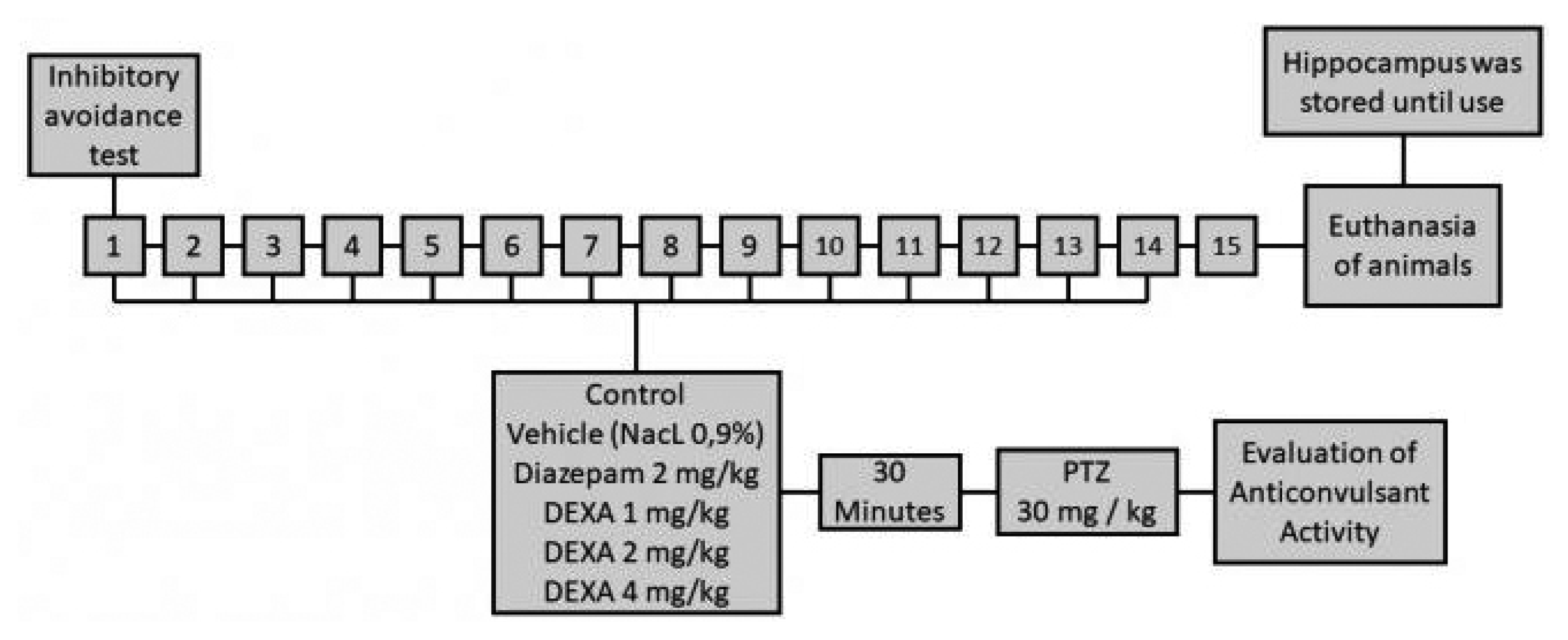
Male Wistar rats were selected from the central vivarium at Universidade Federal do Rio Grande do Sul at 8–9 weeks of age (250–300 g). The animals were handled under standard laboratory conditions consisting of 12 hours light and 12 hours dark cycle and fixed temperature (22–24°C), with free access to food and water. They were divided into six experimental groups (n=9–10 animals per group) that were treated for a period of 14 days on alternate days with group 1, 2 and 3 being the controls, where group 1 is the control group that received no treatment, group 2 that received saline (vehicle of the tested drugs) and group 3 that received diazepam (2 mg/kg), an anticonvulsant drug that is usually one of the first to be used in patients with epilepsy. Group 4, 5 and 6 were treated with the drug dexamethasone, steroidal anti-inflammatory drug, in different concentrations (1, 2 and 4 mg/kg, respectively). The animals in group 1 did not receive treatments and were considered the baseline group for the analysis of OS.
Fig. 1 is a schematic figure explaining experimental protocol.
Evaluation of anticonvulsant activity
The kindling model, considered a chronic model of epilepsy, was used.
5 In this model, the animals received the same doses as described for the treatments for 14 days and, on alternate days, also received subconvulsant doses of pentylenetetrazole (PTZ) intraperitoneally (30 mg/kg). PTZ is a pro-convulsant agent, used to mimic experimental models of seizure
in vivo, which acts through the inhibition of gabaergic receptors, thus promoting neural hyperexcitation by blocking the influx of Cl− and K+
6 efflux. PTZ was injected 30 minutes after the treatments were administered. In these groups, the latency time for the onset of the epileptic seizure (of any kind or intensity) and animal mortality was observed. In a previous study, we have already published the scores presented by the animals according to the Racine scale.
7
Assessment of memory
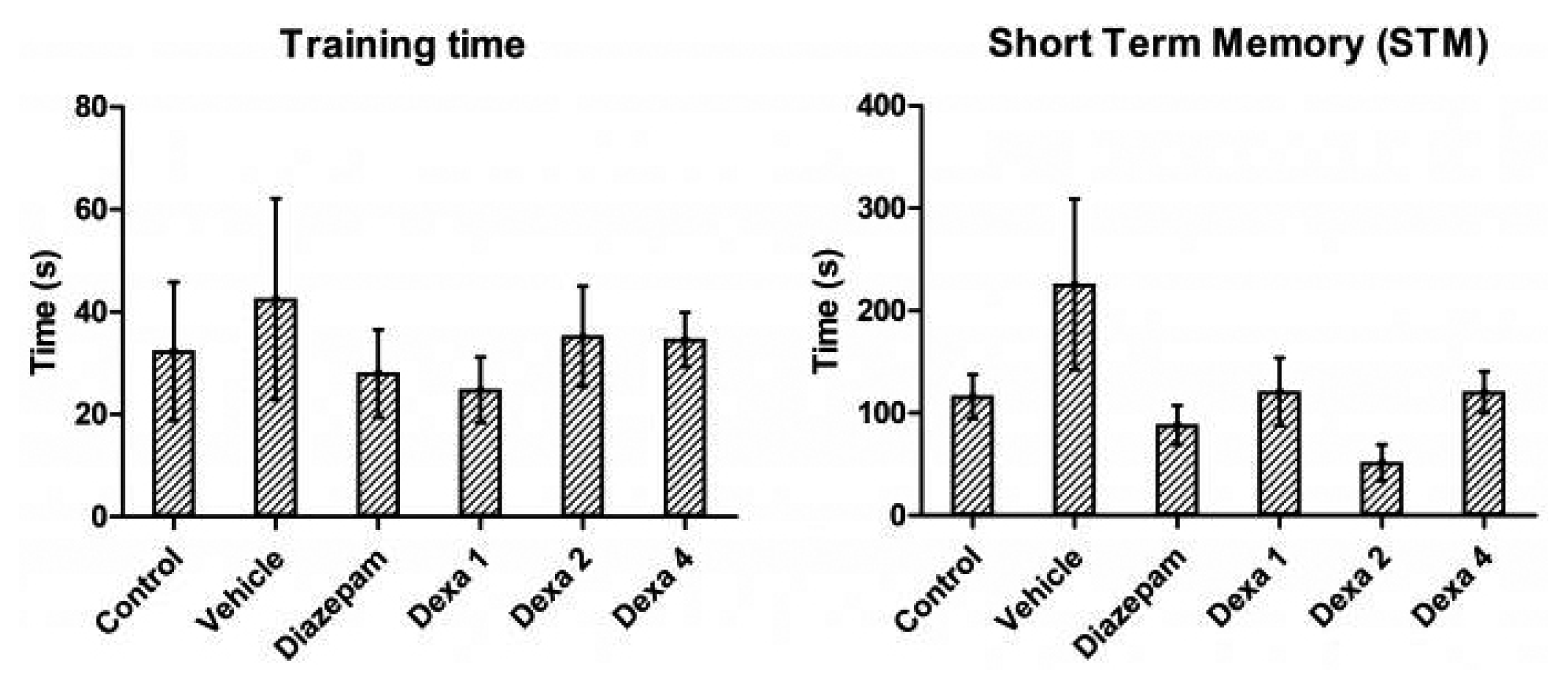
In order to assess the work memory (short term memory, STM), the inhibitory avoidance test was used. The animals are placed on a platform (2.5 cm high by 8.0 cm wide) in an acrylic box with dimensions of 50×25×25 cm. The floor of this box consists of a box of parallel steel bars, with a distance of 0.2 cm between these bars. The time it takes for the animal to descend the platform and place the four legs on the floor of this box was counted. At this point, the animal received a shock in its 0.5 mA paws for 2 seconds. This first moment is called the training section.
8 The time it takes the animal to descend from the platform the first time was not used for any analysis or any kind of segregation. Immediately after the training session, the animals received the pharmacological treatments evaluated via i.p. Two hours after the training session, the animals were placed on the platform again and the time it takes the animal to make the new descent to the ground was counted. At this time the animal did not receive a new electrical discharge. With this test it was possible to check the STM.
8
Evaluation of antioxidant activity
After the end of the anticonvulsant evaluation, the animals were sacrificed by decapitation and the hippocampus was removed to assess the antioxidant activity in the control and treated groups. The samples were stored in a freezer −80°C until the tests were performed. The evaluation of antioxidant activity was carried out through the tests of lipid peroxidation, sulfhydryl groups, activity of the enzymes superoxide dismutase (SOD), catalase and glutathione peroxidase. In these evaluations, 5 to 8 animals were used per group.
Oxidative damage
Lipid damage was determined by the method based on thiobarbituric acid reactive substances (TBARS), which is widely used as a sensitive method for measuring lipid peroxidation, described by Wills.
9 The results are expressed in nmol TBARS/mg protein. The content of sulfhydryl groups, a measure of non-enzymatic cell defense, was determined through its reduction with 5,5′-dithiobis-2-nitrobenzoic acid (Ellman's reagent) producing a colored compound, read spectrophotometrically at 412 nm.
9
Total proteins
The determination of total proteins was carried out by the method of Lowry
10 and the values were expressed in mg/mL. This method has the advantage of its high sensitivity, being used for the determination of proteins in different tissues.
10
Superoxide dismutase activity
The activity of the SOD enzyme was measured spectrophotometrically in all samples as described by Bannister and Calabrese.
11 SOD can be measured indirectly, following the decrease in absorbance of oxidized adrenaline at 480 nm. The results were expressed in U SOD/mg protein. One unit is defined as the amount of enzyme that inhibits the speed of adenochrome formation by 50%.
11
Catalase activity
The measurement method for catalase activity (CAT) was that described by AEBI,
12 in which the enzyme catalyzes the decomposition of hydrogen peroxide into H
2O and O
2. The decomposition speed of H
2O
2 was measured spectrophotometrically at 230 nm for 180 seconds. The results were expressed in mmol H
2O
2/mg protein/minute.
12
Glutathione peroxidase activity
The activity of glutathione peroxidase (GPX) was described by Paglia,
12 in which the measurement occurred through changes in the absorbance of nicotinamide adenine dinucleotide phosphate (NADPH) at 340 nm. The results were expressed in nmol NADPH/mg protein/minute.
Statistical analysis
The data were presented as mean and standard error. After defining the subgroups, statistical analysis was performed using analysis of variance followed by the Tukey post-hoc test. Results with p<0.05 were considered statistically significant (F-values are presented only if p<0.05). All statistical analyzes were performed using a database that was assembled in the SPSS version 17 statistical package (IBM Corp., Armonk, NY, USA).
Ethical considerations
In compliance with Law No. 11,794/2008, chapter IV, art.14, §4, the number of animals to be used for the execution of a project and the duration of each experiment were the minimum necessary to produce the conclusive result, saving to the maximum, the suffering animal. Ethical procedures for the care and use of animals were adopted according to the regulations published by the Brazilian Society for Neuroscience and Behavior. The present study is part of a larger project called “influence of inflammation on the epileptogenic process” that was approved by the Health Sciences Research Committee and the Ethics Committee on the use of animals at UFRGS on 12/18/2012.
Discussion
The excess of positive charges promotes depolarization of the neuronal membrane, which causes voltage-dependent calcium channels to open; calcium, in turn, is able to activate intracellular enzymes responsible for triggering harmful processes, in addition to activating the enzyme phospholipase A2 (responsible for the metabolism of membrane lipids, which will trigger an inflammatory response) and mobilizing synaptic vesicles, thus triggering the synaptic processes that will generate neuronal hyperexcitability. This fine line between OS and neuroinflammation, predisposing to seizures, has already been well established. With the understanding of this mechanism, there is the possibility of using anti-inflammatory drugs as an alternative therapy for the treatment and in certain refractory epilepsies. It is strongly believed that OS has a great influence on the occurrence of seizures.
13–
15 The animals' ability to memorize was tested in one moment. Steroid-specific receptors are known to be largely concentrated in the hippocampus, septal area and amygdala parts of the brain intimately involved in behavior, mood, learning and memory. Despite this, dexamethasone, a steroidal anti-inflammatory drug, acting on steroid receptors, has no deleterious effect on working memory in doses tested.
16
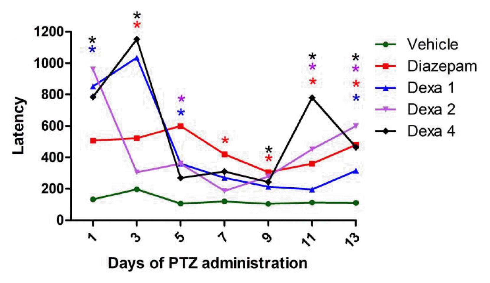
Continuing the behavioral analysis, the latency time for the first seizure was compared in the groups that received PTZ. It can be seen that the vehicle group, which did not receive any pharmacological treatment, had the shortest latency times as compared to the Diazepam control group and to all DEXA groups. Among the groups tested, there were no statistical differences between the mean latency times, allowing to state that DEXA, in all dosages, had an effect similar to diazepam in increasing latency for the first seizure. It has been seen in a model similar to this study that turmeric
16 was able to increase the latency time for the first seizure due to its anti-inflammatory and antioxidant effects. On the other hand, neuroprotective compounds such as yerba mate
17 and grape juice
18 were not able to produce this effect. In another animal model of epilepsy, the pilocarpine model, a similar result was found. The dose of DEXA used was 10 mg/kg and no one anticonvulsive drug was used as a control. There was a reduction in the latency time for the beginning of seizure demonstrating the possible anticonvulsant effect of DEXA on the animal model of pilocarpine.
19
As already mentioned, in a previous study we have already published the scores presented by the animals according to the Racine scale; however, the analysis of the latency for the onset of conclusive crises is of utmost importance for understanding the mechanism of action of this drug. It was possible to verify that DEXA acts in reducing not only the intensity of seizures, but also the onset time for seizures, reinforcing the possible anticonvulsant and neuroprotective role of this drug.
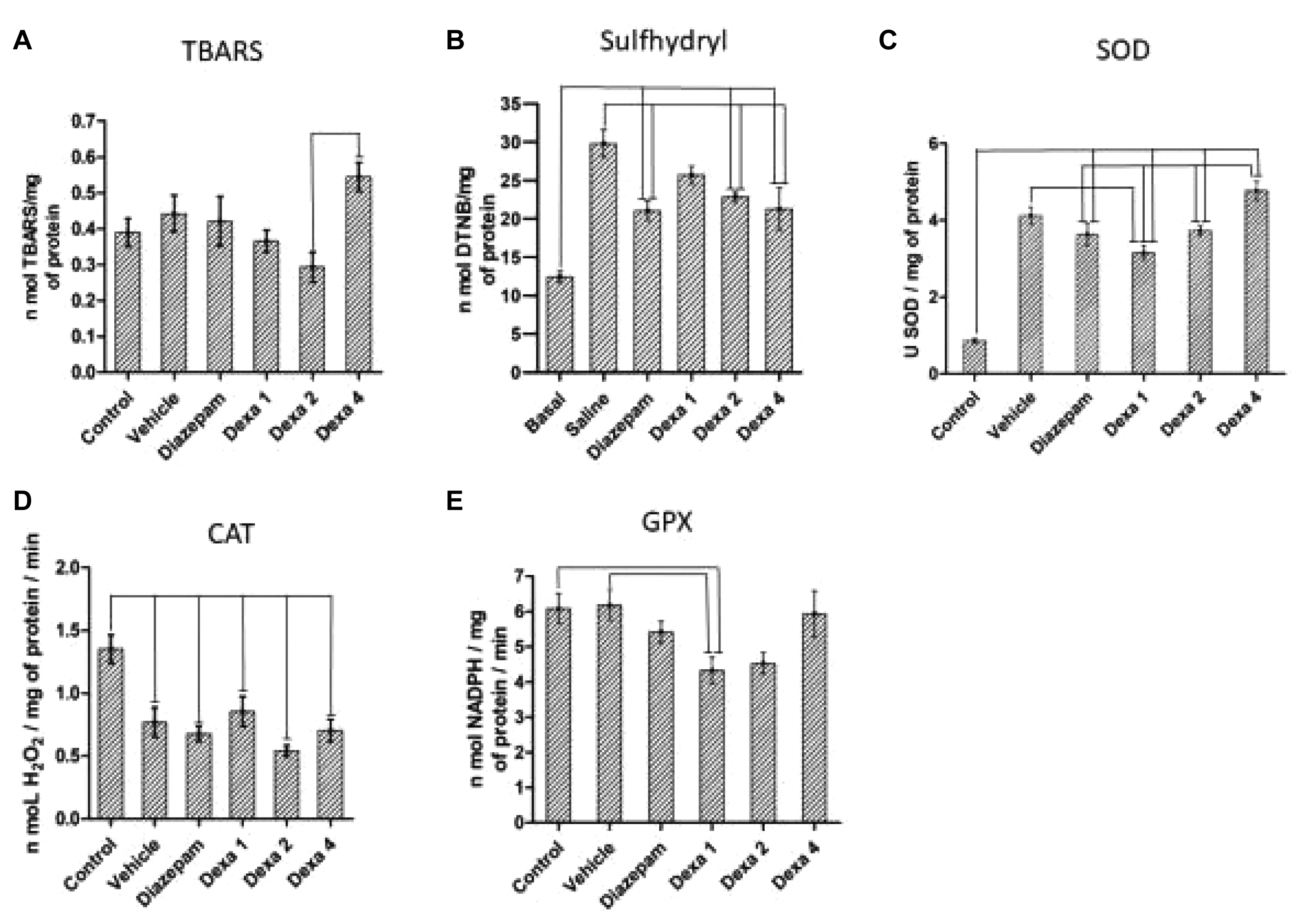
7 As for OS, no group differed from the control groups (vehicle and diazepam) in relation to TBARS levels; however, an increase in lipid peroxidation was found in the DEXA group 4 mg/kg compared to the DEXA 2 mg/kg group. The SOD enzyme catalyzes the dismutation of superoxide into oxygen and hydrogen peroxide and because of this, it is an important antioxidant defense. The increase in this enzyme in all groups treated with PTZ compared to the baseline group, found in this study, is an unexpected result. The use of PTZ generated an increase in SOD. This increase was seen in all groups that received PTZ. Neither the use of diazepam nor DEXA at any concentration was able to reverse this increase and return levels to baseline values. A trend towards a reduction in SOD levels was seen with the use of DEXA at the lowest concentration, whereas at the highest dose, the levels remained as high as in the saline group. Unlike the result found in this study, Qi et al.
20 (2018) found decreased levels of SOD related to PTZ treatment in the brain of rats. This difference can be explained by the difference in the experimental design since in the mentioned study there was only one administration of PTZ and in the present study the animals received PTZ for 2 weeks. This increase is probably generated by the chronic use of PTZ. It is noteworthy that the mentioned study evaluated the total brain tissue, and the present study the hippocampus. The increase found in the groups that received DEXA can be explained by the OS-inducing effect of this drug.
21
As for sulfhydryl levels, an increase was found in all treated groups when compared to the baseline group. This increase, caused by PTZ, was attenuated in the groups that received diazepam and DEXA at the two highest doses; however, there was no return to baseline values. In the DEXA 1 group, there was an increase compared to baseline, but it remained similar to the vehicle group, demonstrating that DEXA at this dose did not attenuate the OS caused by PTZ administration. There was a decrease in the CAT enzyme caused by the administration of PTZ. This decrease was not reversed by the use of DEXA or diazepam. Other studies demonstrate an increase in CAT related to PTZ administration both in the hippocampus
22 and in all brain tissue.
21,
22 Reversal of this effect was found by other drugs such as metformin (first-line therapy for patients with type 2 diabetes since it has antioxidant, anti-inflammatory and neuroprotective properties)
20 and by compounds such as pycnogenol (Pinus pinaster bark extract, which contains flavonoids and has antioxidant, anti-inflammatory, neuroprotective and cardio protective properties),
21 the peony extract (extracted from the root barks, which has antioxidant and anti-inflammatory effects)
22 and the yerba mate (
Ilex paraguariensis, anti-inflammatory effects).
17 Regarding the activity of the GPX enzyme, there was a tendency to decrease the activity with the use of DEXA, and the dose of 1 mg generated a significant reduction compared to the vehicle and control groups. In the other DEXA concentrations and diazepam-treated group, there were no significant differences compared to the baseline and control groups.
Compensatory mechanisms can explain the results obtained, comparing the groups in which PTZ doses were administered in relation to the baseline group. As the enzyme activity of SOD increases, H2O2 can accumulate in the tissue. This accumulation could be metabolized by the enzymatic action of GPX and CAT, but there was little difference in the levels of GPX and decreased activity of CAT. In this situation, there would be a notable increase in H2O2 levels, which could (for example by the Fenton reaction) generate a high degree of hydroxyl ion concentration. However, the OS marker used (TBARS) showed no difference between the groups. Sulfhydryl groups were high compared to baseline. This may mean a non-enzymatic compensatory mechanism on redox signaling since the enzyme system was flawed, but there was no increase in TBARS levels among all groups. The anticonvulsant effect of DEXA remains uncertain. Immunological mechanisms, with the release of cytokines and inflammatory mediators, seem to be the key to this process. Mechanisms that generate OS may also be related to the observed effect. New studies are needed to investigate a new therapeutic approach in the treatment of epilepsy with this drug.


